 |
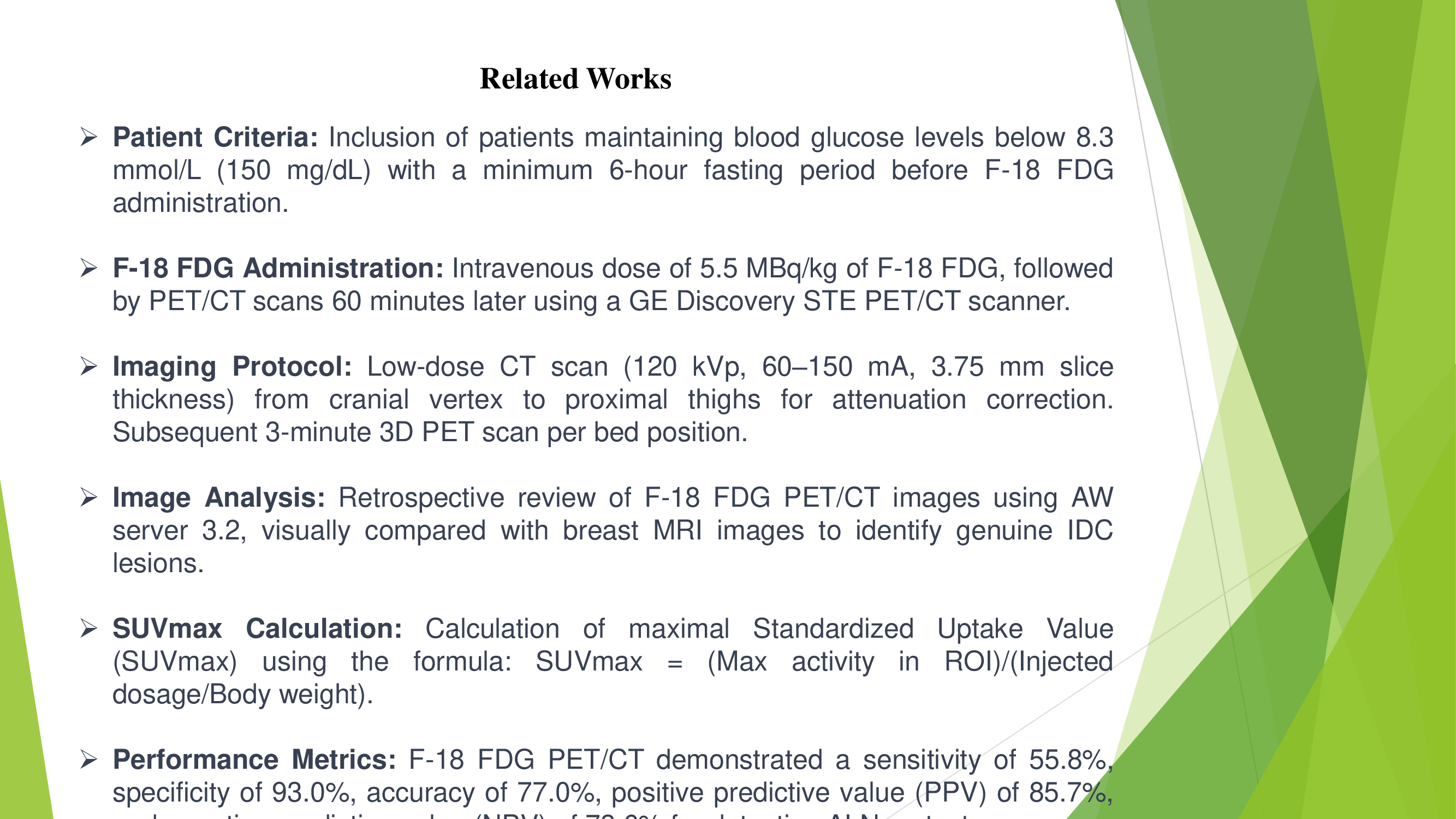
This slide outlines the methodology employed in related works, specifically focusing on patient criteria, F-18 FDG administration, imaging protocol, image analysis, SUVmax calculation, and performance metrics. Patient inclusion involved maintaining blood glucose levels below 8.3 mmol/L with a minimum fasting period. F-18 FDG was administered intravenously, followed by PET/CT scans using a GE Discovery STE PET/CT scanner. The imaging protocol included a low-dose CT scan for attenuation correction and subsequent 3D PET scans. Image analysis retrospectively reviewed F-18 FDG PET/CT images, visually compared with breast MRI images to identify genuine IDC lesions. The maximal Standardized Uptake Value (SUVmax) was calculated using a specific formula. Performance metrics revealed F-18 FDG PET/CT sensitivity of 55.8%, specificity of 93.0%, accuracy of 77.0%, positive predictive value (PPV) of 85.7%, and negative predictive value (NPV) of 73.6% for detecting axillary lymph node metastases. |
 IEEE/ICACT20240384 Slide.07
[Big Slide]