 IEEE/ICACT20250100 Slide.13
[Big Slide]
Oral Presentation
| Virual Oral Presentation Slide by Slide |
|
IEEE/ICACT20250100 Slide.13
[Big Slide]
| Oral Presentation |
|
|
|
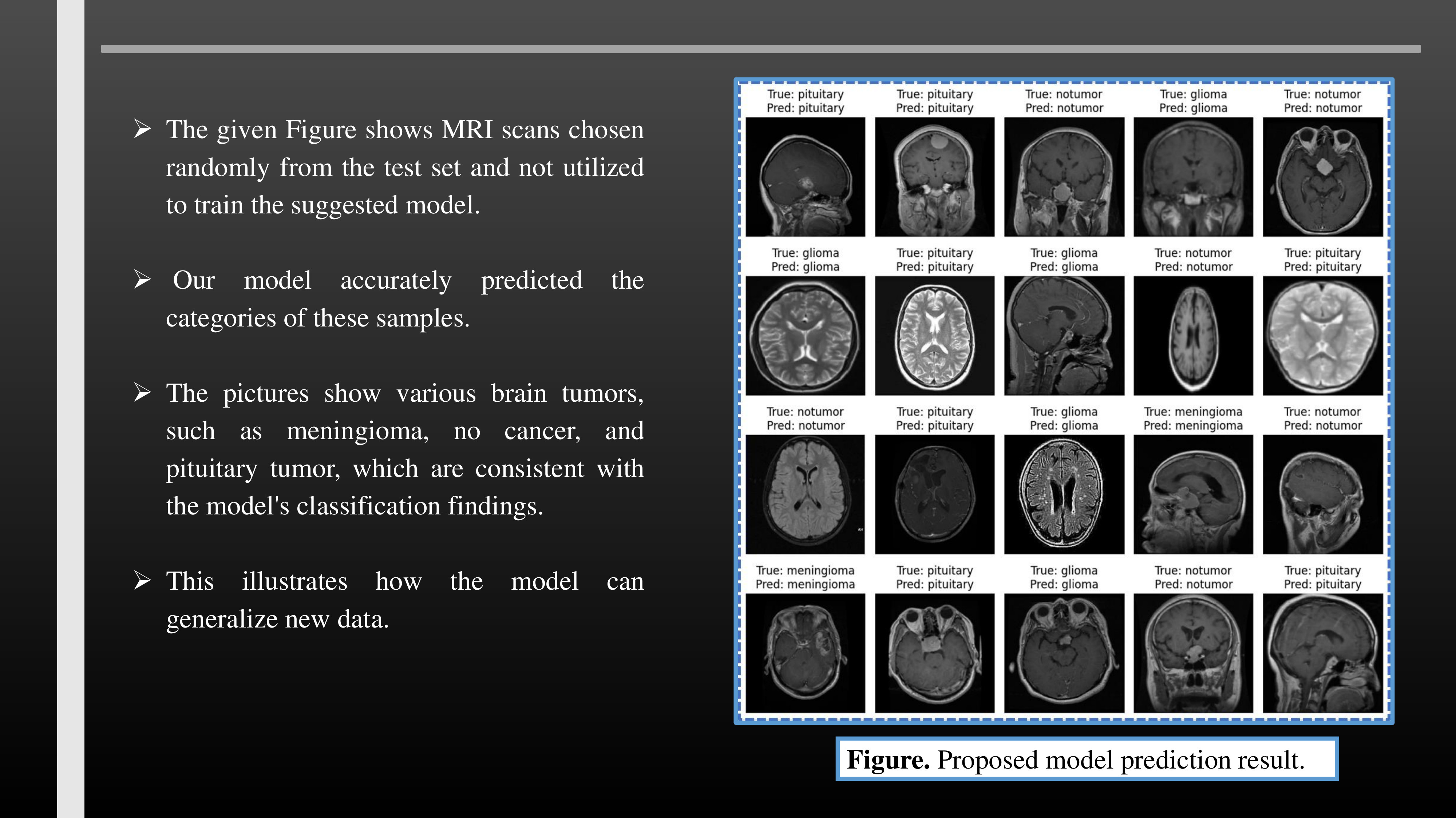
The given Figure shows MRI scans chosen randomly from the test set and not utilized to train the suggested model. Our model accurately predicted the categories of these samples. The pictures show various brain tumors, such as meningioma, no cancer, and pituitary tumor, which are consistent with the model's classification findings. This illustrates how the model can generalize new data. | |
| [Go to Next Slide] |