ICACT20240327 Slide.16
[Big slide for presentation] |
Chrome Text-to-Speach Click!! |
 |
As we conclude our presentation, I extend my sincere gratitude for your time and attention. Thank you for being a part of this discussion.
|
|
ICACT20240327 Slide.15
[Big slide for presentation]
|
Chrome Text-to-Speach Click!! |
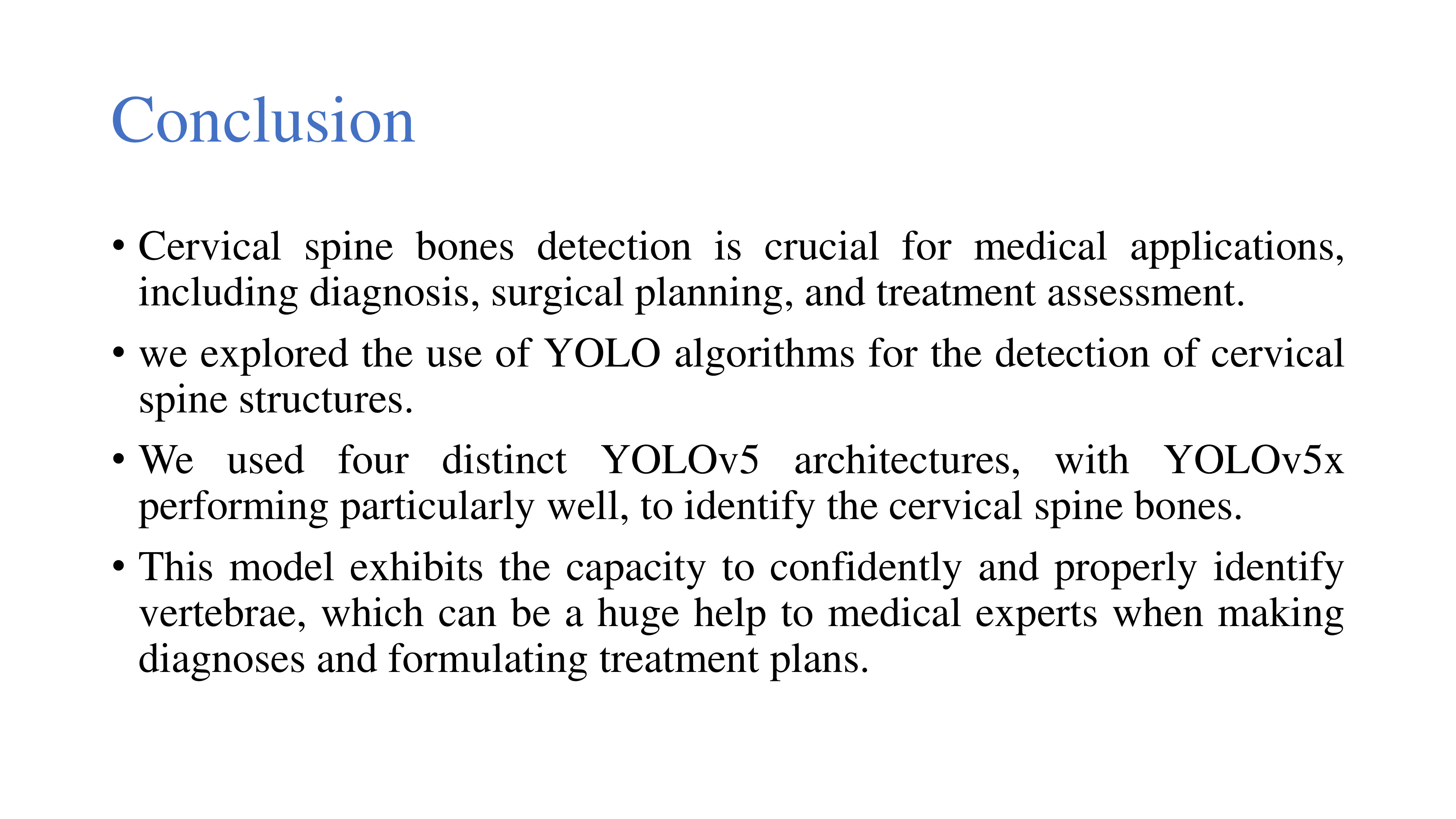 |
As we arrive at the 'Conclusion' of our study, it is evident that cervical spine bones detection holds paramount significance in diverse medical applications, ranging from diagnosis to surgical planning and treatment assessment.
Throughout our exploration, we harnessed the power of YOLO algorithms for the precise detection of cervical spine structures. Utilizing four distinct YOLOv5 architectures, our comparative analysis revealed the outstanding performance of YOLOv5x. This particular model demonstrated exceptional accuracy in identifying cervical spine bones.
The robust capabilities of our model extend a helping hand to medical experts, providing them with a reliable tool for confident and accurate vertebrae identification. This, in turn, stands to significantly impact the diagnostic process and treatment planning within the medical field. In conclusion, our research underscores the promising potential of YOLO algorithms, particularly YOLOv5x, in advancing cervical spine bone detection for improved healthcare outcomes.
|
|
ICACT20240327 Slide.14
[Big slide for presentation]
|
Chrome Text-to-Speach Click!! |
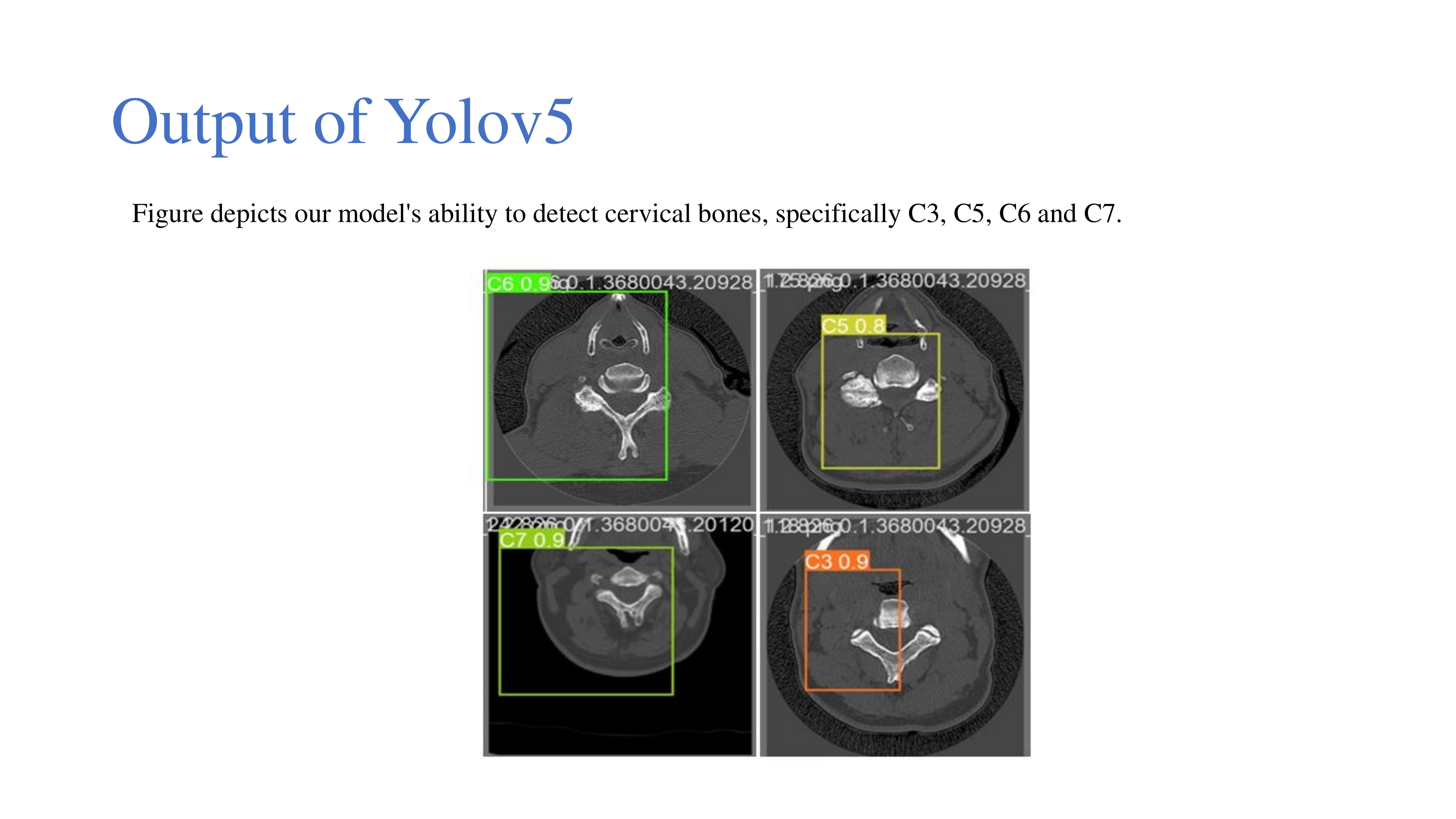 |
Moving forward, let's explore the 'Output of YOLOv5.' In this figure, our model's effectiveness in detecting cervical bones is vividly illustrated. The results showcase the remarkable accuracy of our system, with specific instances highlighted:
C3 detected with a confidence of 90%,
C5 detected with a confidence of 80%,
C6 detected with a confidence of 90%, and
C7 detected with a confidence of 90%.
This visual representation serves as a testament to the robustness and precision of our YOLOv5 model in successfully identifying individual cervical bones, reinforcing the efficacy of our approach in cervical spine bone detection.
|
|
ICACT20240327 Slide.13
[Big slide for presentation]
|
Chrome Text-to-Speach Click!! |
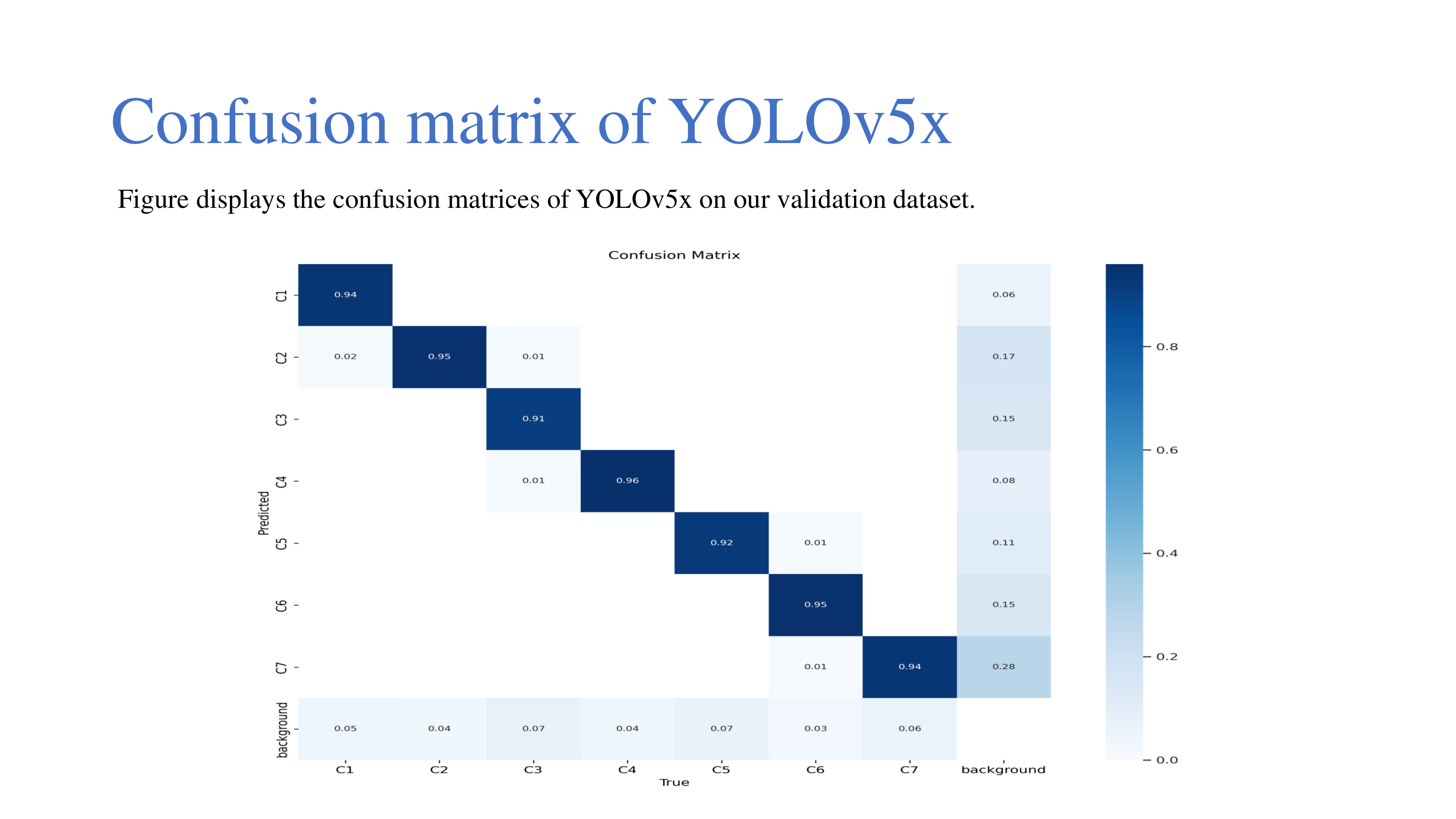 |
Turning our attention to the 'Confusion Matrix of YOLOv5x,' this figure provides a comprehensive visual representation of the model's performance on our validation dataset. The confusion matrix offers a detailed breakdown, showcasing the accuracy of detecting cervical bones C1, C2, C3, C4, C5, C6, and C7.
This analytical tool allows us to examine not only the correct classifications but also instances of misclassifications, aiding in the identification of potential areas for improvement. By scrutinizing the confusion matrix, we gain valuable insights into the model's proficiency in accurately identifying each cervical bone, contributing to a thorough assessment of the YOLOv5x performance in the context of our specific application.
|
|
ICACT20240327 Slide.12
[Big slide for presentation]
|
Chrome Text-to-Speach Click!! |
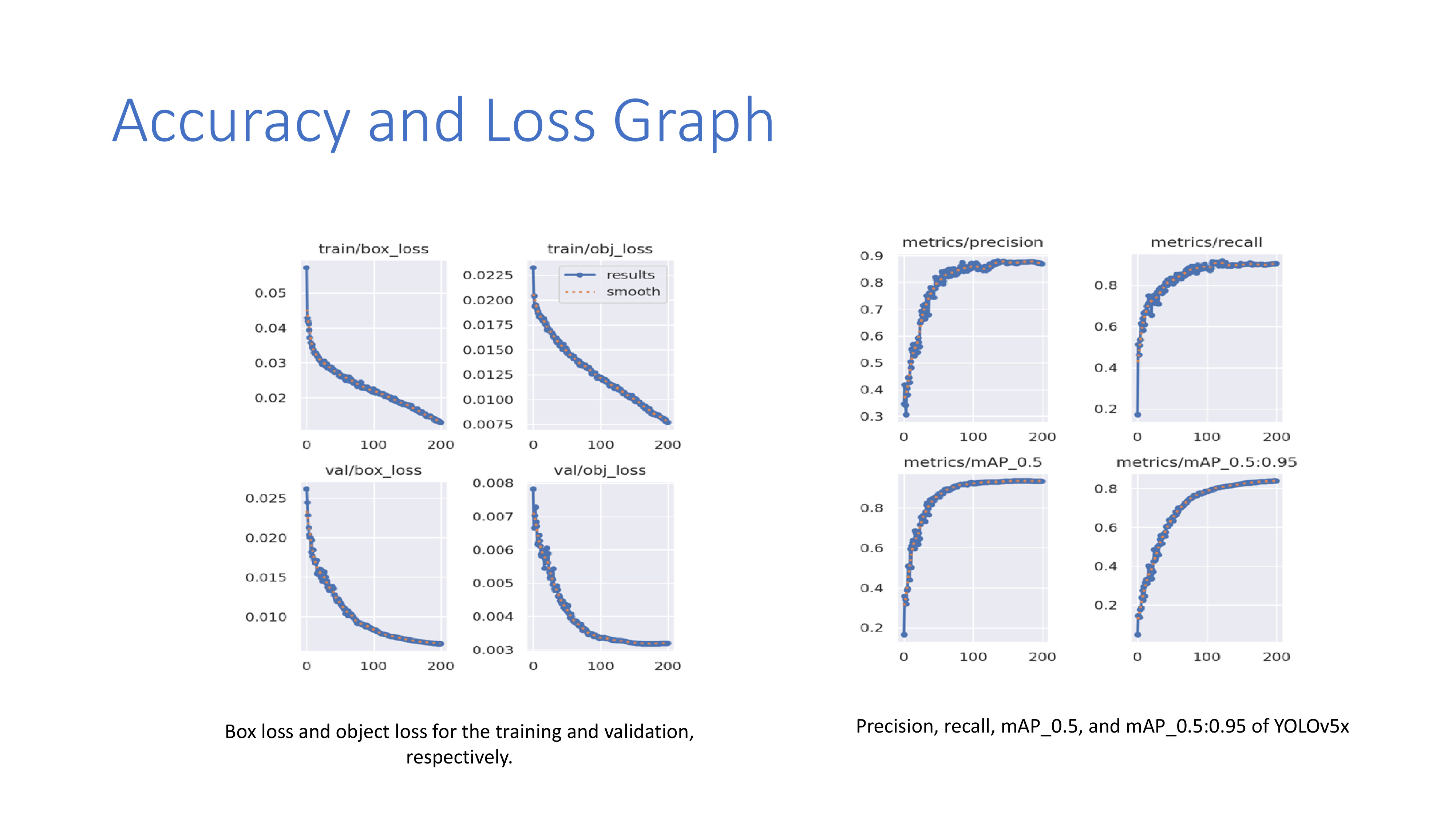 |
Now, let's delve into the 'Accuracy and Loss Graph' to gain deeper insights into our model's performance metrics. The graph showcases the box loss and object loss for both the training and validation phases, providing a visual representation of the learning dynamics.
Additionally, we'll examine key metrics, including precision, recall, mAP_0.5, and mAP_0.5:0.95 specifically for YOLOv5x. These metrics offer a comprehensive evaluation of the model's accuracy, sensitivity, and performance across various thresholds.
By analyzing these crucial indicators, we gain a nuanced understanding of the strengths and potential areas for improvement in our YOLOv5x model, shaping the discussion around the overall efficacy of our cervical spine bone detection system.
|
|
ICACT20240327 Slide.11
[Big slide for presentation]
|
Chrome Text-to-Speach Click!! |
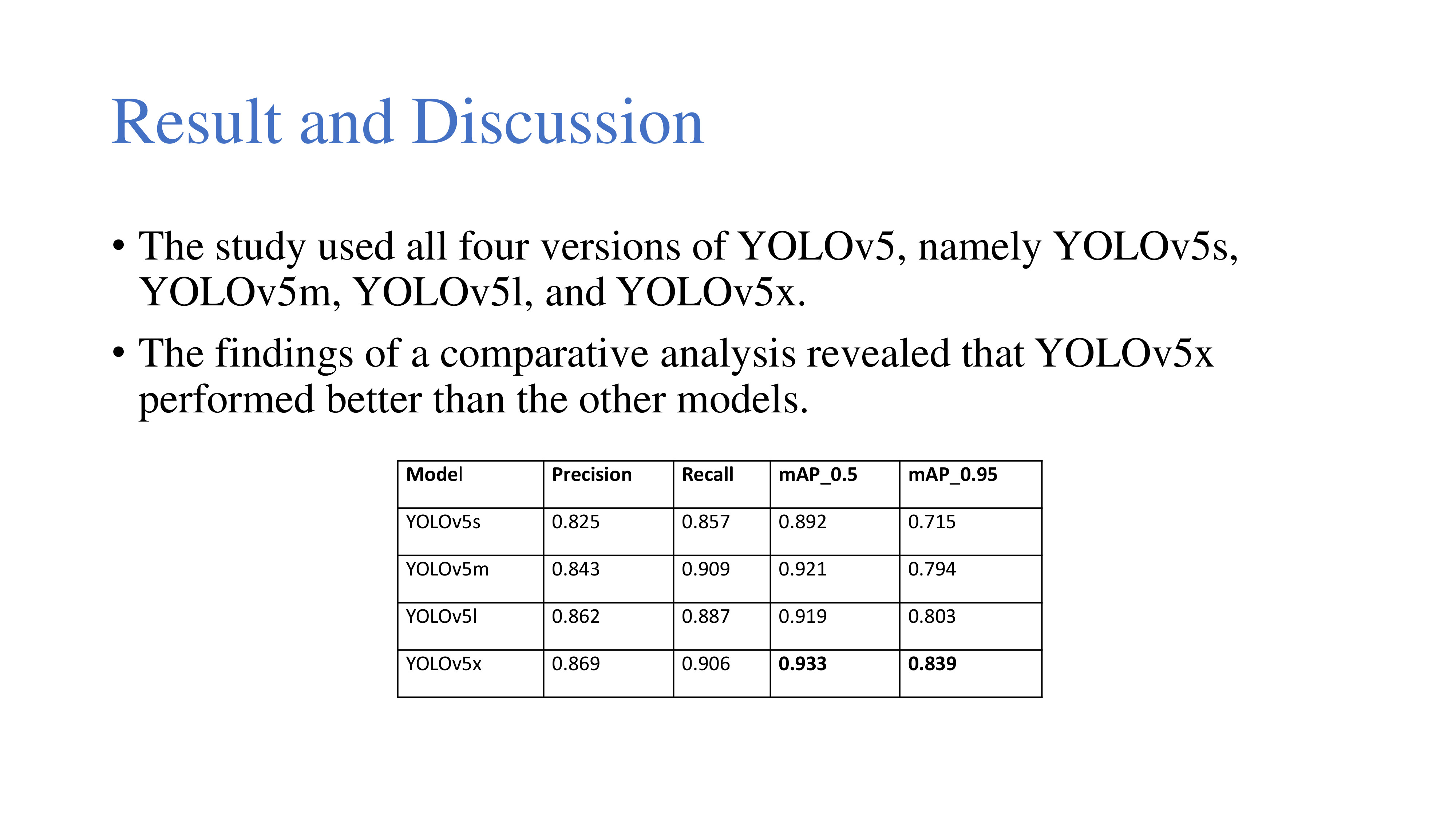 |
Transitioning to the pivotal stage of our study – 'Result and Discussion.' In our exploration, all four versions of YOLOv5, including YOLOv5s, YOLOv5m, YOLOv5l, and YOLOv5x, were rigorously employed.
Through a careful comparative analysis, the findings illuminated a distinct performance superiority of YOLOv5x over its counterparts. The precision, efficiency, and overall effectiveness of YOLOv5x in our specific application surpassed that of other models, underscoring its significance in achieving superior results for cervical spine bone detection.
|
|
ICACT20240327 Slide.10
[Big slide for presentation]
|
Chrome Text-to-Speach Click!! |
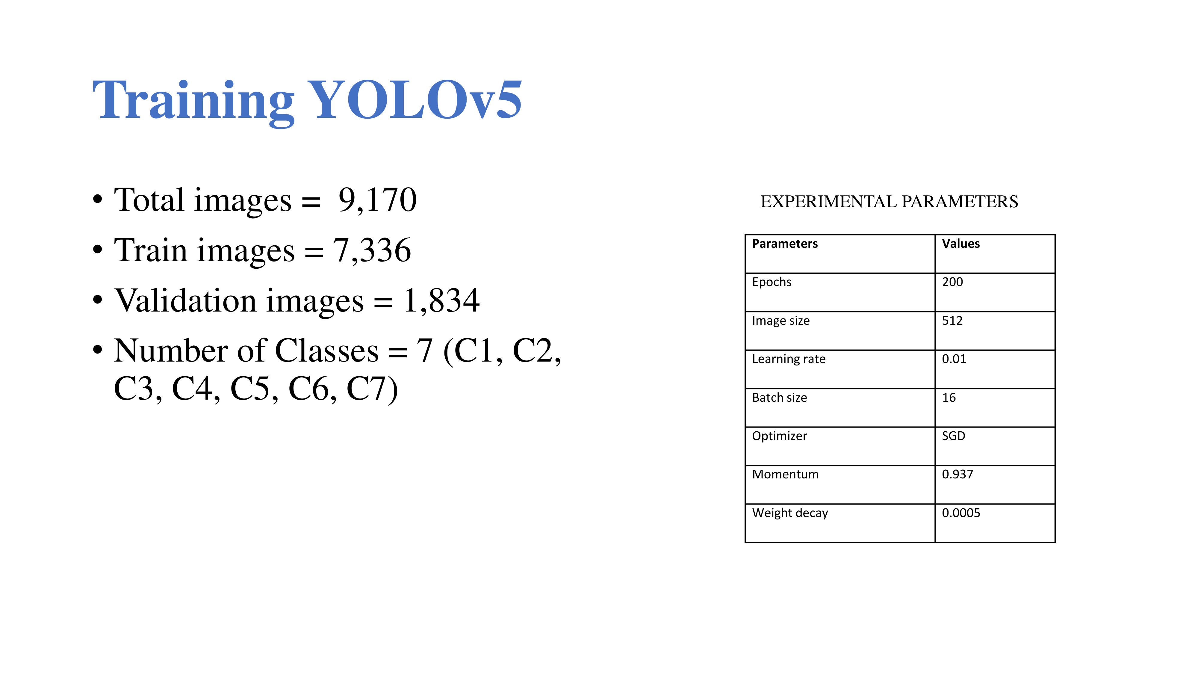 |
Now, let's dive into the details of our 'Training YOLOv5' phase. Our dataset, comprising a total of 9,170 images, was divided into 7,336 for training and 1,834 for validation. These images represent the anatomical regions corresponding to vertebrae C1 to C7.
For the training process, we set up the following experimental parameters:
Epochs: 200
Image Size: 512
Learning Rate: 0.01
Batch Size: 16
Optimizer: SGD (Stochastic Gradient Descent)
Momentum: 0.937
Weight Decay: 0.0005
These carefully chosen parameters aim to optimize the YOLOv5 model, ensuring effective learning and accurate detection of cervical spine bones. As we progress through the training phase, these settings play a crucial role in fine-tuning the model for our specific application.
|
|
ICACT20240327 Slide.09
[Big slide for presentation]
|
Chrome Text-to-Speach Click!! |
 |
Now, let's explore the cornerstone of our methodology – 'YOLOv5.' YOLOv5, which stands for 'You Only Look Once' version 5, is an open-source object detection framework. Uniquely designed for real-time scenarios, YOLOv5 provides a compelling blend of speed and accuracy, making it versatile for a wide range of applications.
This framework introduces a streamlined architecture, marked by enhanced performance when compared to its predecessors, solidifying its status as a popular choice for computer vision tasks. YOLOv5's appeal lies in its commitment to simplicity, efficiency, and state-of-the-art performance, garnering significant recognition within the machine learning and computer vision communities. As we integrate YOLOv5 into our study, its capabilities become instrumental in achieving precise and efficient cervical spine bone detection.
|
|
ICACT20240327 Slide.08
[Big slide for presentation]
|
Chrome Text-to-Speach Click!! |
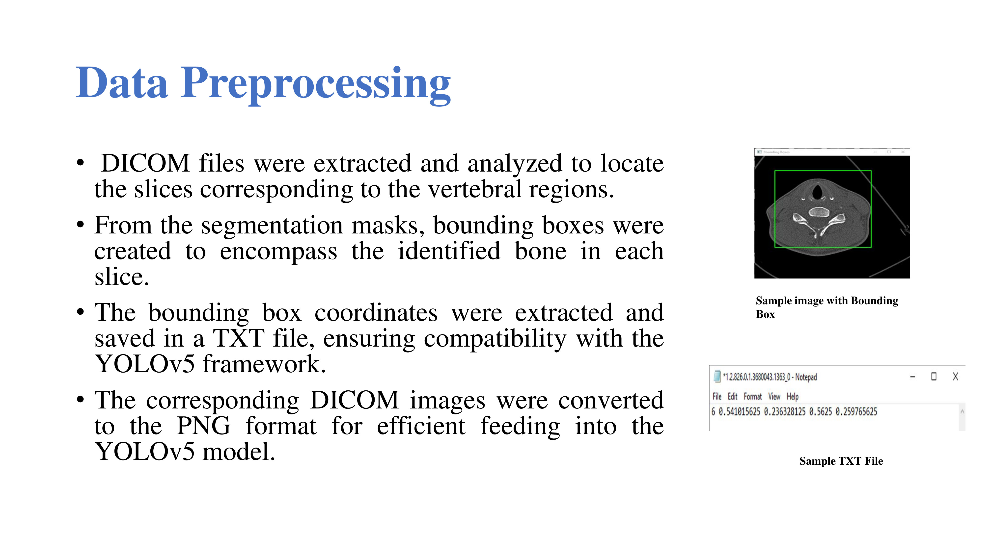 |
Now, let's look at the details of our "Data Preprocessing" phase.
The first step involved extracting and carefully analyzing DICOM files to identify the slices that corresponded to the relevant vertebral regions. Next, based on the segmentation masks, bounding boxes were skillfully generated to encapsulate the identified bones within each slice.
To seamlessly integrate this information into the YOLOv5 framework, the bounding box coordinates were extracted and thoughtfully saved in a TXT file, ensuring compatibility with the YOLOv5 model. Additionally, for enhanced efficiency in model feeding, the corresponding DICOM images underwent conversion to the PNG format.
This comprehensive data preprocessing pipeline lays the foundation for accurate and effective utilization of our dataset in training the YOLOv5 model for cervical spine bone detection.
|
|
ICACT20240327 Slide.07
[Big slide for presentation]
|
Chrome Text-to-Speach Click!! |
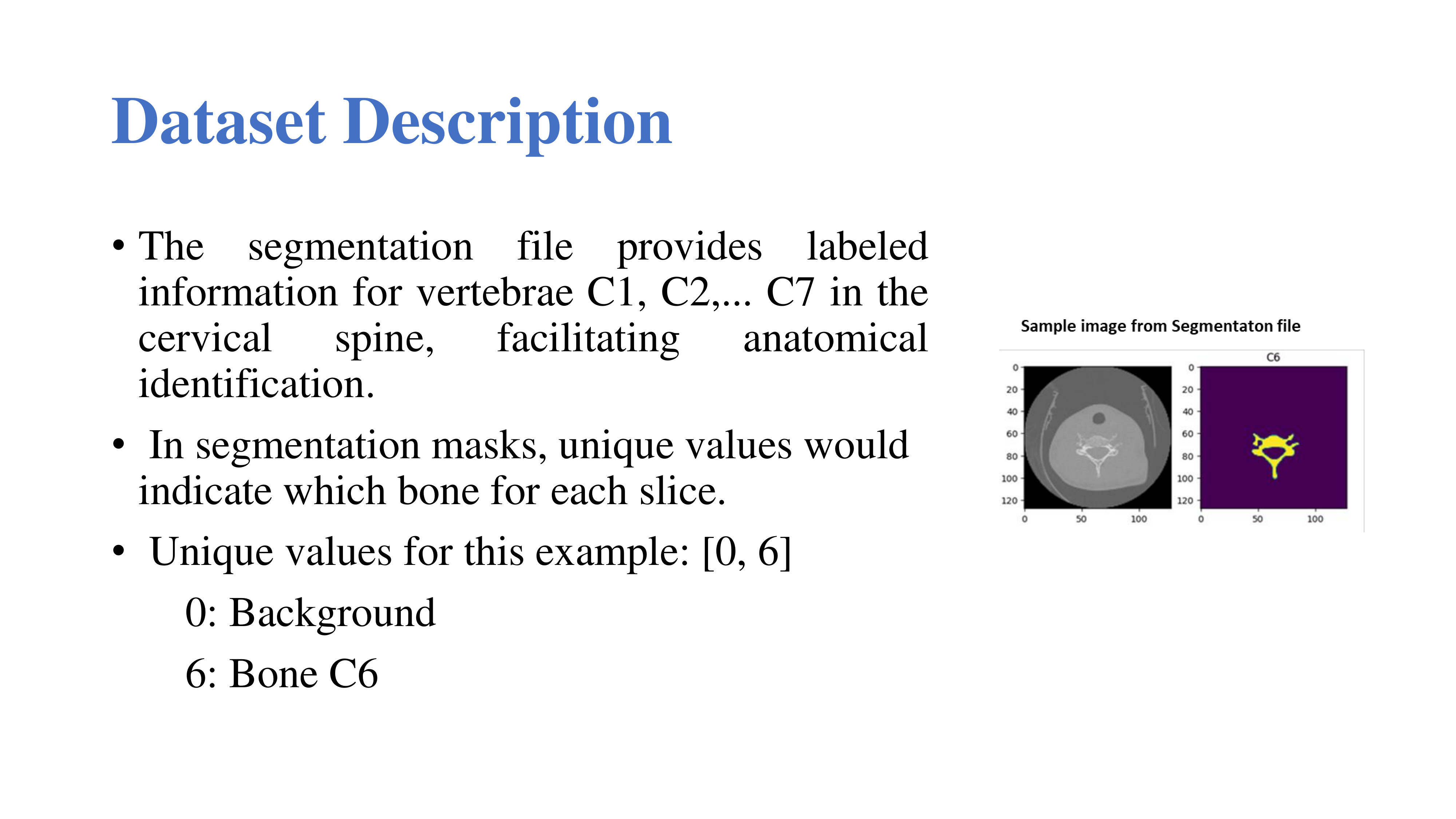 |
Now, let's get into the specifics of our dataset, with a focus on the 'Dataset Description' field. The segmentation file within our dataset proves instrumental, providing labeled information for each vertebra in the cervical spine, from C1 to C7. This segmentation allows for the precise anatomical identification of each vertebra.
In the segmentation masks, unique values play a key role in indicating the presence of specific bones for each slice. For instance, in our example, we have unique values [0,6]. Here's the breakdown:
0: Represents the background
6: Specifically denotes Bone C6
This specific level of labeling not only speeds up model training but also ensures accurate and detailed identification of cervical spine bones throughout the dataset.
|
|
ICACT20240327 Slide.06
[Big slide for presentation]
|
Chrome Text-to-Speach Click!! |
 |
Moving on to our dataset, which forms the backbone of our study. The data utilized in this research is sourced from the RSNA 2022 Cervical Spine Fracture Detection competition hosted on Kaggle. Within this dataset, three main folders hold invaluable information: 'train images,' 'Bounding box,' and 'segmentations.' Each subfolder is dedicated to a specific patient, containing multiple slice images pertinent to the corresponding case. This comprehensive dataset serves as the foundation for training and evaluating our model, providing a diverse range of medical images for a thorough investigation into cervical spine bone detection.
|
|
ICACT20240327 Slide.05
[Big slide for presentation]
|
Chrome Text-to-Speach Click!! |
 |
Turning our attention to the 'Need for Accurate and Efficient Cervical Spine Bones Detection Method,' we emphasize the critical importance of precise detection methods in ensuring timely and appropriate treatment while minimizing the risk of complications. Delays or missed diagnoses can lead to inadequate treatment, prolonged recovery times, and even irreversible neurological damage. Traditional methods, such as visual inspection of radiographic images by human experts, are not only time-consuming but also susceptible to human error.
Recognizing these challenges, deep learning-based methods emerge as a promising solution. These methods have the potential to significantly enhance the speed and accuracy of cervical spine bone detection, enabling earlier diagnosis and treatment. By leveraging advanced technologies, we aim to address the pressing need for efficient detection methods that can positively impact patient outcomes and overall healthcare quality.
|
|
ICACT20240327 Slide.04
[Big slide for presentation]
|
Chrome Text-to-Speach Click!! |
 |
Exploring the 'Significance of Detecting Cervical Spine Bones in Medical Applications,' we underscore the critical role of accurate detection in diagnosing and treating injuries and medical conditions affecting this region. In the realm of medical applications, the detection of cervical spine bones proves indispensable for assessing traumatic injuries like fractures or dislocations. Moreover, its precision holds immense importance in the planning and execution of surgical procedures, particularly in the context of cervical spine surgery. Beyond treatment, the accurate detection of cervical bones contributes significantly to medical education, enhancing the understanding of the cervical spine's structure and function for students and healthcare professionals alike.
|
|
ICACT20240327 Slide.03
[Big slide for presentation]
|
Chrome Text-to-Speach Click!! |
 |
In this slide, we introduce the intricate details of the cervical spine, which constitutes the uppermost section of the spine. Comprising seven vertebrae labeled as C1 through C7, these structures play a vital role in supporting the head and neck. Beyond its structural significance, the cervical spine holds a pivotal role in facilitating the flexibility and range of motion necessary for the movement of our head in various directions. Moreover, it serves as a protective shield for the spinal cord, an essential component of our nervous system responsible for transmitting signals to and from the brain. The significance of the cervical spine becomes evident when considering that injuries or certain medical conditions affecting it can lead to symptoms such as pain, numbness, or weakness in the neck, arms, or hands.
|
|
ICACT20240327 Slide.02
[Big slide for presentation]
|
Chrome Text-to-Speach Click!! |
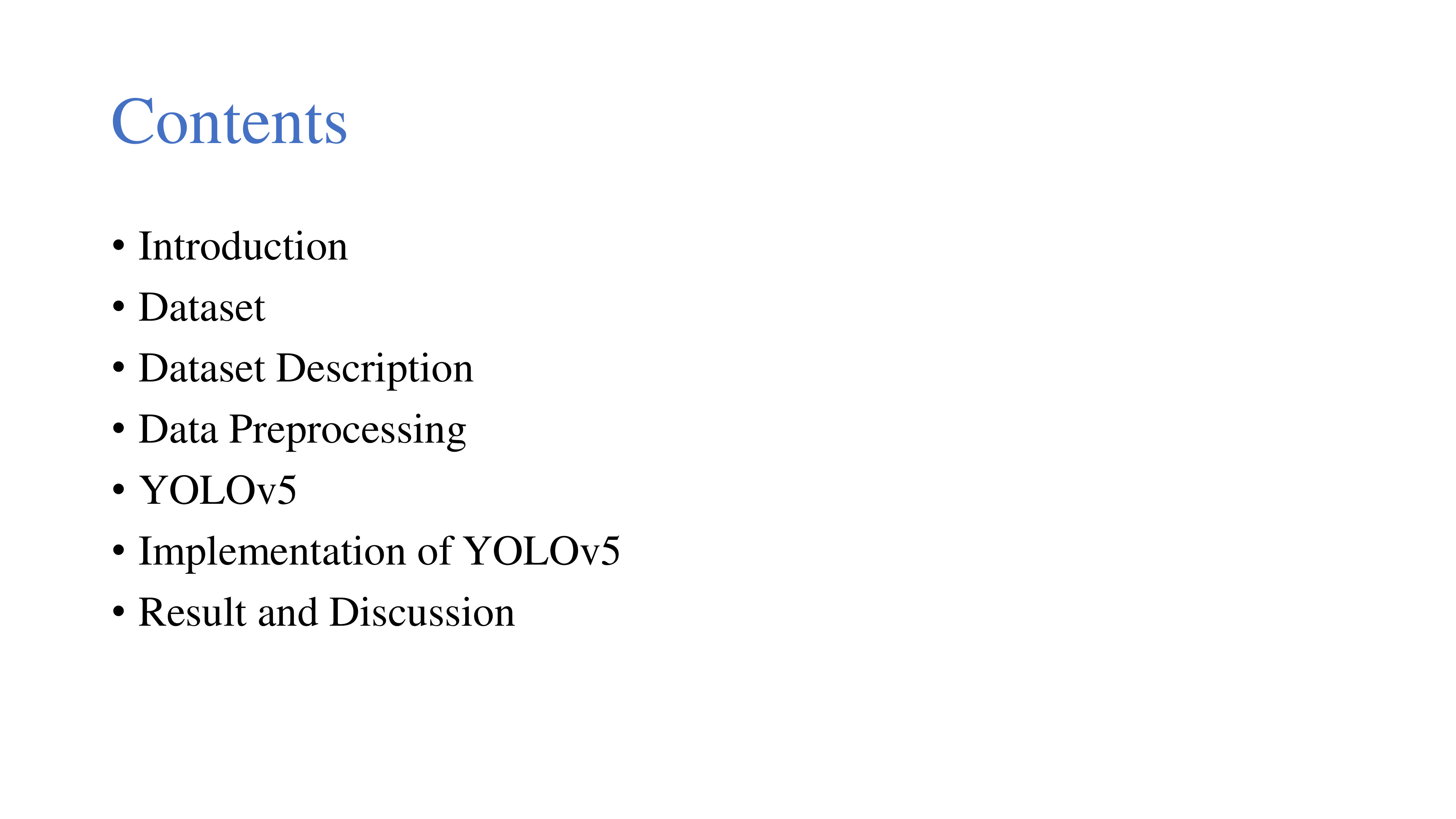 |
Here are the contents of the presentation. The presentation will commence with an introduction, providing an overview of the topic. Following that, we'll take a look at the dataset and its description., outlining key aspects of its origin and composition. Subsequently, we'll explore the data preprocessing steps undertaken to prepare our dataset. Moving on, we'll focus on YOLOv5, the chosen deep learning model, and discuss its implementation in our study. Finally, we'll conclude by presenting the results and engaging in a discussion of our findings.
|
|
ICACT20240327 Slide.01
[Big slide for presentation]
|
Chrome Text-to-Speach Click!! |
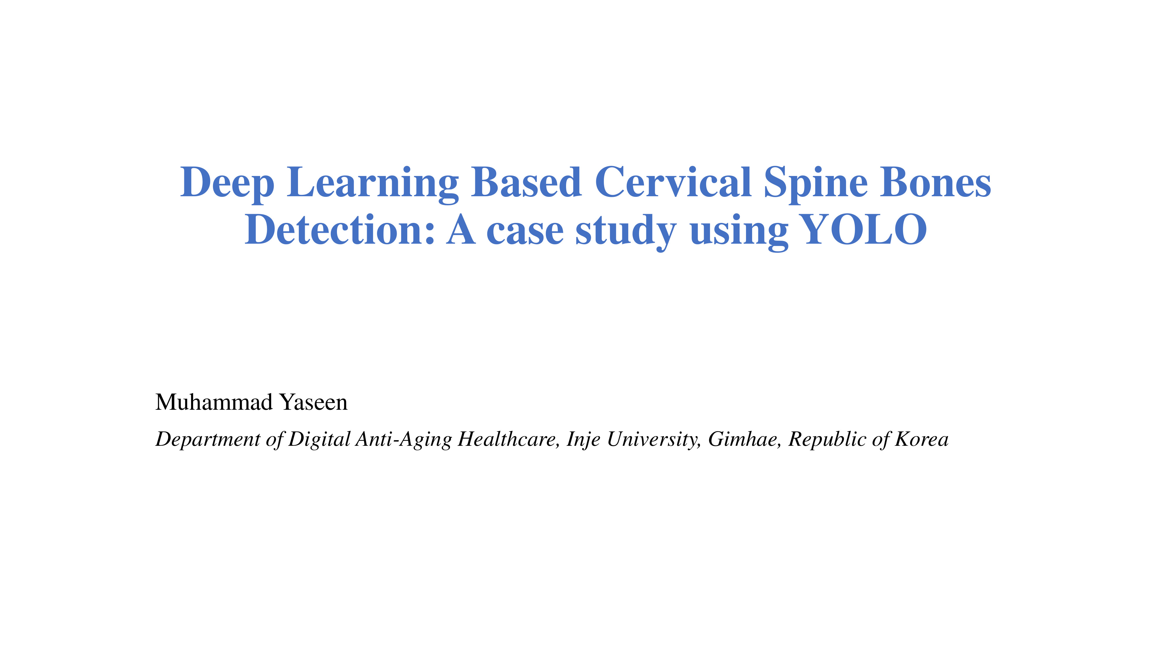 |
Hi everyone! My name is Muhammad Yaseen, and I'm from the Department of Digital Anti-Aging Healthcare at Inje University in South Korea. Today, I am thrilled to present our research on “Deep Learning Based Cervical Spine Bones Detection: A Case Study using YOLO.”
The core of our investigation revolves around the groundbreaking application of deep learning techniques, specifically leveraging the YOLO (You Only Look Once) algorithm, for the precise detection of cervical spine bones.
|
|
ICACT20240327 Slide.00
[Big slide for presentation]
|
Chrome Text-to-Speach Click!! |
 |
|