ICACT20250100 Slide.16
[Big slide for presentation]
[YouTube] |
Chrome Text-to-Speach Click!! |
 |
If you have any questions, ask them in the YouTube video comments
|
|
ICACT20250100 Slide.15
[Big slide for presentation]
|
Chrome Text-to-Speach Click!! |
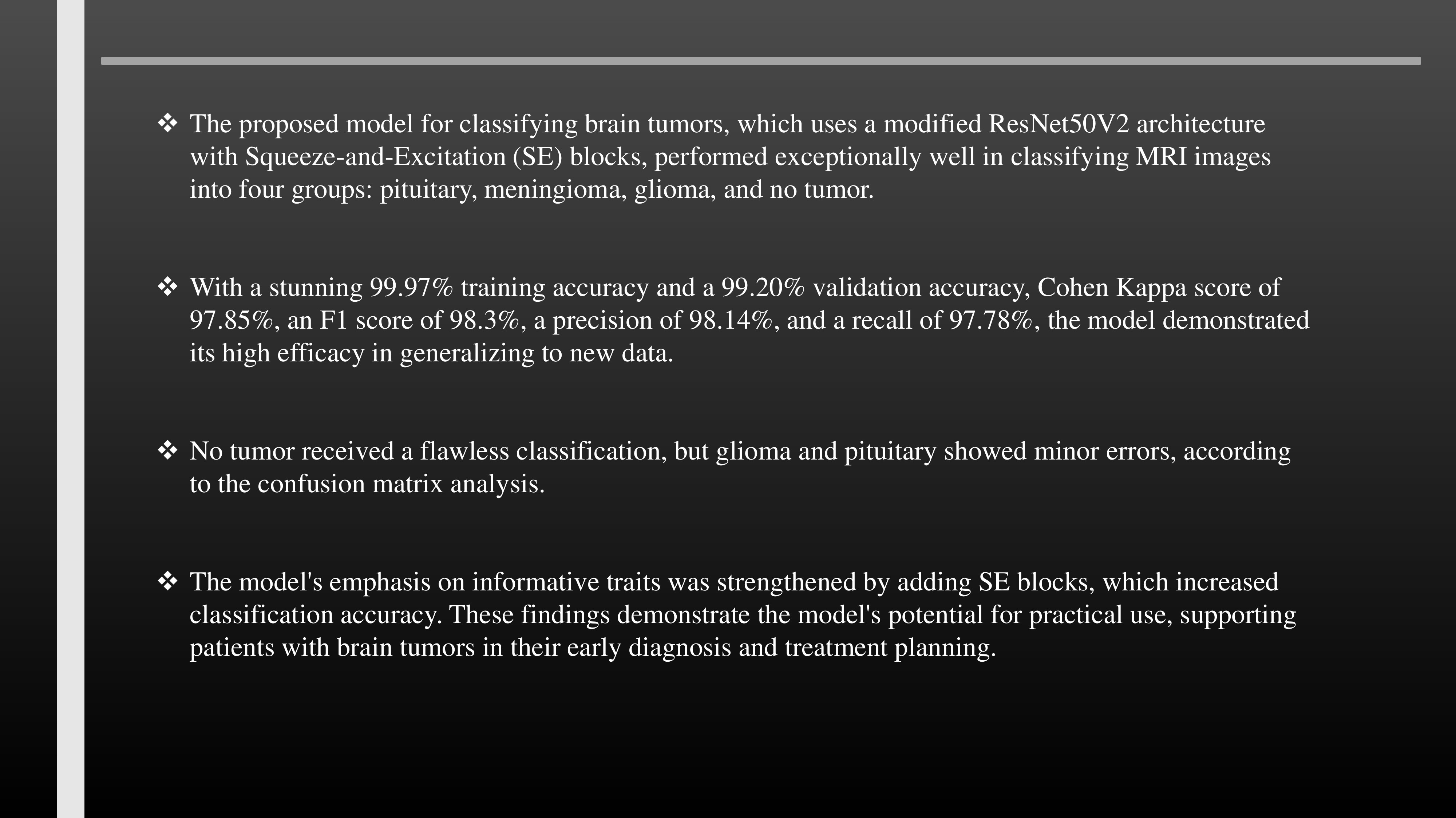 |
The proposed model for classifying brain tumors, which uses a modified ResNet50V2 architecture with Squeeze-and-Excitation (SE) blocks, performed exceptionally well in classifying MRI images into four groups: pituitary, meningioma, glioma, and no tumor.
With a stunning 99.97% training accuracy and a 99.20% validation accuracy, Cohen Kappa score of 97.85%, an F1 score of 98.3%, a precision of 98.14%, and a recall of 97.78%, the model demonstrated its high efficacy in generalizing to new data.
No tumor received a flawless classification, but glioma and pituitary showed minor errors, according to the confusion matrix analysis.
The model's emphasis on informative traits was strengthened by adding SE blocks, which increased classification accuracy. These findings demonstrate the model's potential for practical use, supporting patients with brain tumors in their early diagnosis and treatment planning.
|
|
ICACT20250100 Slide.14
[Big slide for presentation]
|
Chrome Text-to-Speach Click!! |
 |
4. CONCLUSION
|
|
ICACT20250100 Slide.13
[Big slide for presentation]
|
Chrome Text-to-Speach Click!! |
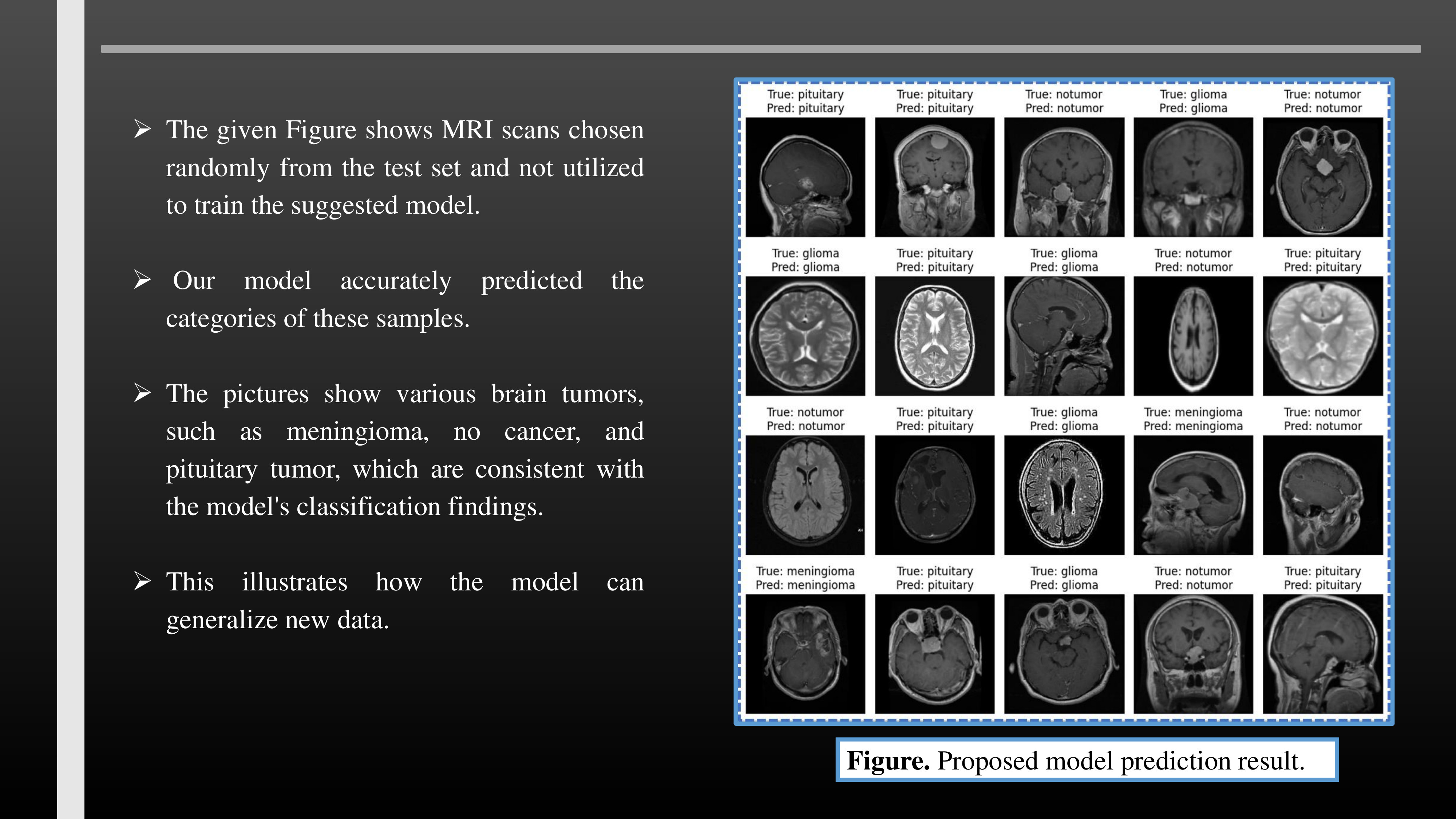 |
The given Figure shows MRI scans chosen randomly from the test set and not utilized to train the suggested model.
Our model accurately predicted the categories of these samples.
The pictures show various brain tumors, such as meningioma, no cancer, and pituitary tumor, which are consistent with the model's classification findings.
This illustrates how the model can generalize new data.
|
|
ICACT20250100 Slide.12
[Big slide for presentation]
|
Chrome Text-to-Speach Click!! |
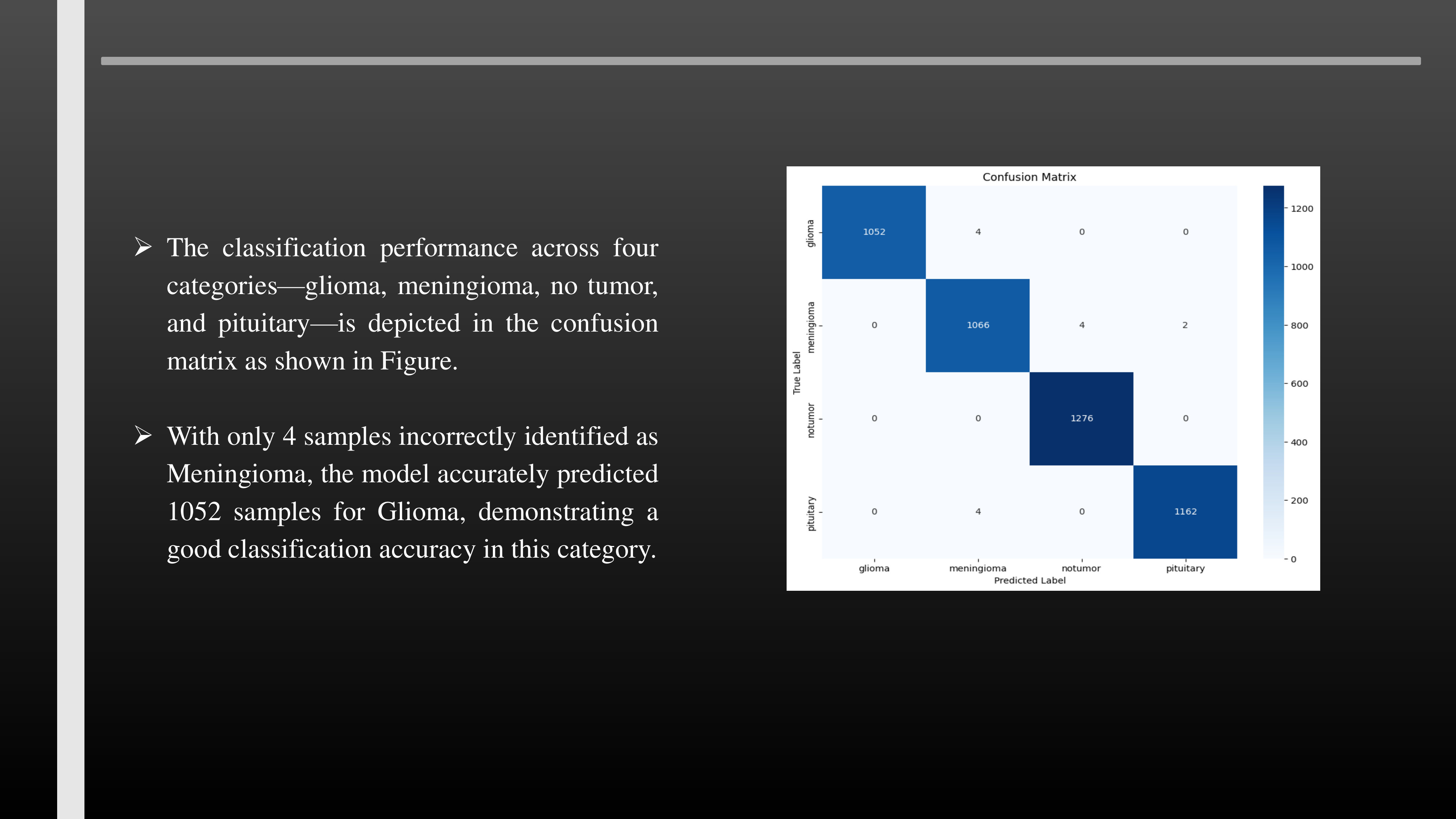 |
The classification performance across four categories—glioma, meningioma, no tumor, and pituitary—is depicted in the confusion matrix as shown in Figure.
With only 4 samples incorrectly identified as Meningioma, the model accurately predicted 1052 samples for Glioma, demonstrating a good classification accuracy in this category.
|
|
ICACT20250100 Slide.11
[Big slide for presentation]
|
Chrome Text-to-Speach Click!! |
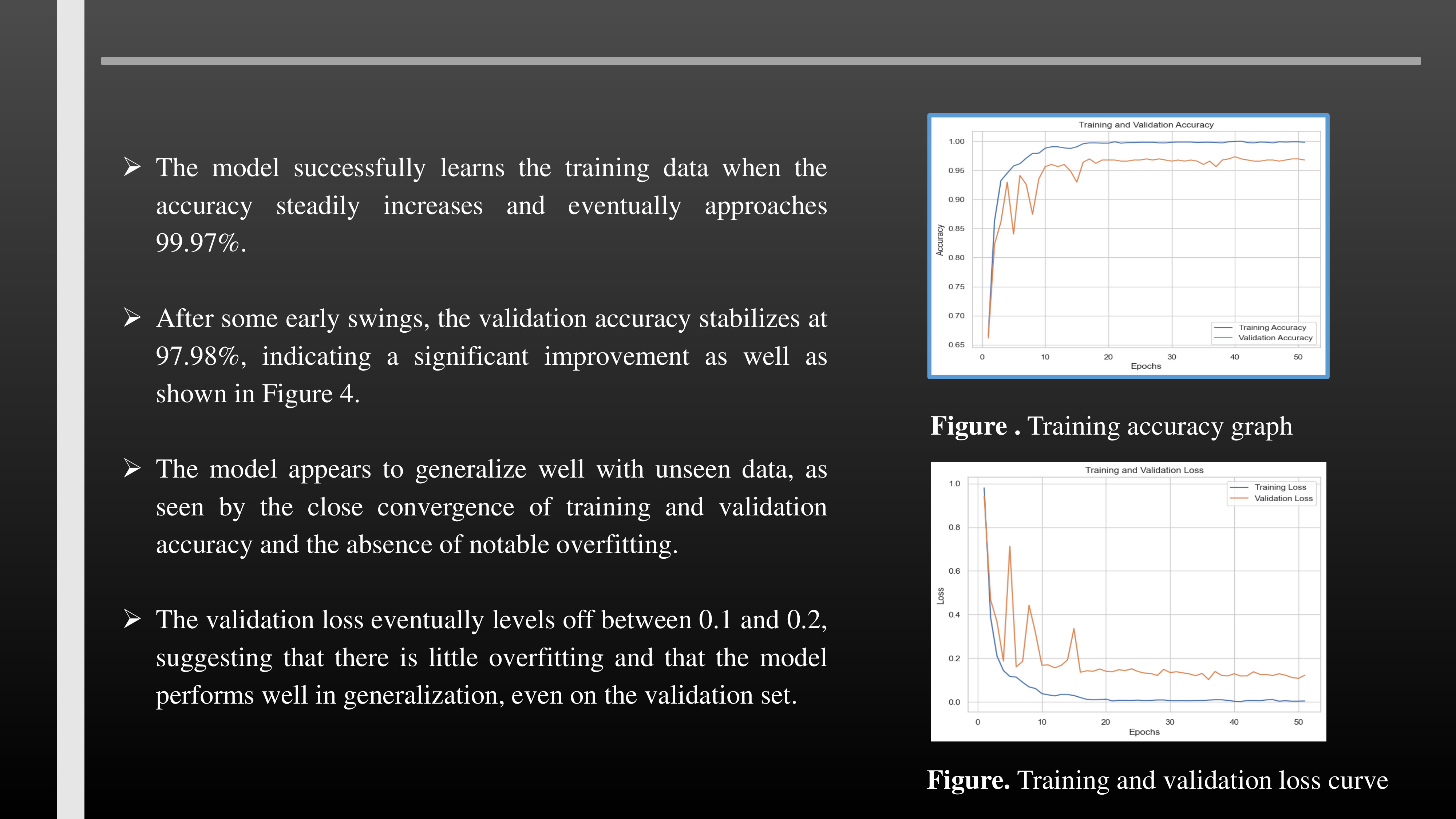 |
The model successfully learns the training data when the accuracy steadily increases and eventually approaches 99.97%.
After some early swings, the validation accuracy stabilizes at 97.98%, indicating a significant improvement as well as shown in Figure 4.
The model appears to generalize well with unseen data, as seen by the close convergence of training and validation accuracy and the absence of notable overfitting.
The validation loss eventually levels off between 0.1 and 0.2, suggesting that there is little overfitting and that the model performs well in generalization, even on the validation set.
|
|
ICACT20250100 Slide.10
[Big slide for presentation]
|
Chrome Text-to-Speach Click!! |
 |
3.RESULTS AND DISCUSSION
|
|
ICACT20250100 Slide.09
[Big slide for presentation]
|
Chrome Text-to-Speach Click!! |
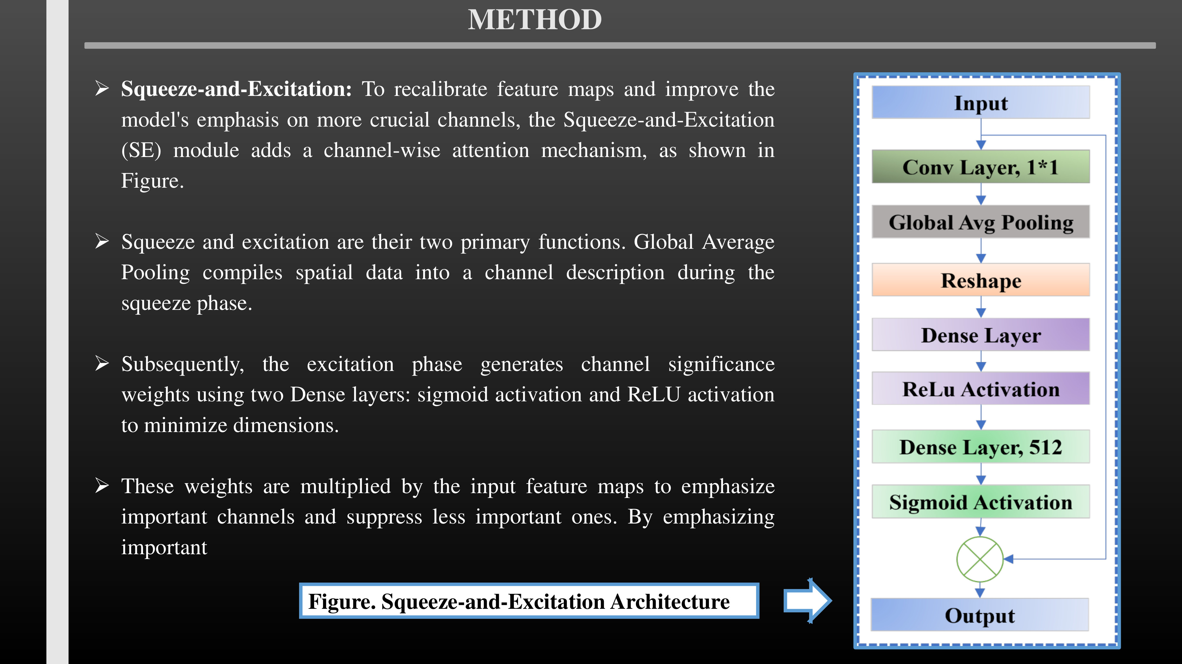 |
Squeeze-and-Excitation: To recalibrate feature maps and improve the model's emphasis on more crucial channels, the Squeeze-and-Excitation (SE) module adds a channel-wise attention mechanism, as shown in Figure.
Squeeze and excitation are their two primary functions. Global Average Pooling compiles spatial data into a channel description during the squeeze phase.
Subsequently, the excitation phase generates channel significance weights using two Dense layers: sigmoid activation and ReLU activation to minimize dimensions.
These weights are multiplied by the input feature maps to emphasize important channels and suppress less important ones. By emphasizing important
|
|
ICACT20250100 Slide.08
[Big slide for presentation]
|
Chrome Text-to-Speach Click!! |
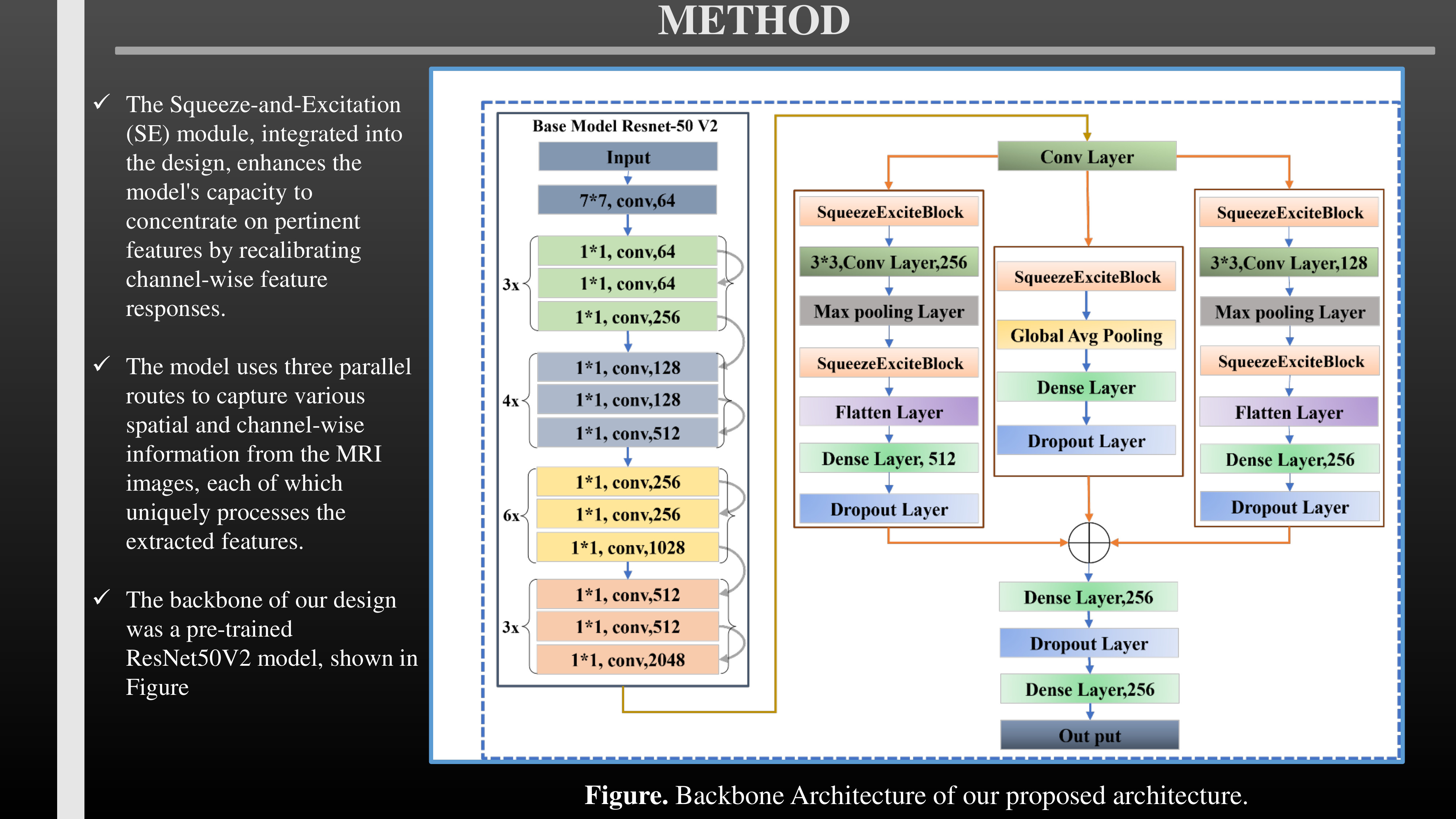 |
The Squeeze-and-Excitation (SE) module, integrated into the design, enhances the model's capacity to concentrate on pertinent features by recalibrating channel-wise feature responses.
The model uses three parallel routes to capture various spatial and channel-wise information from the MRI images, each of which uniquely processes the extracted features.
The backbone of our design was a pre-trained ResNet50V2 model, shown in Figure
|
|
ICACT20250100 Slide.07
[Big slide for presentation]
|
Chrome Text-to-Speach Click!! |
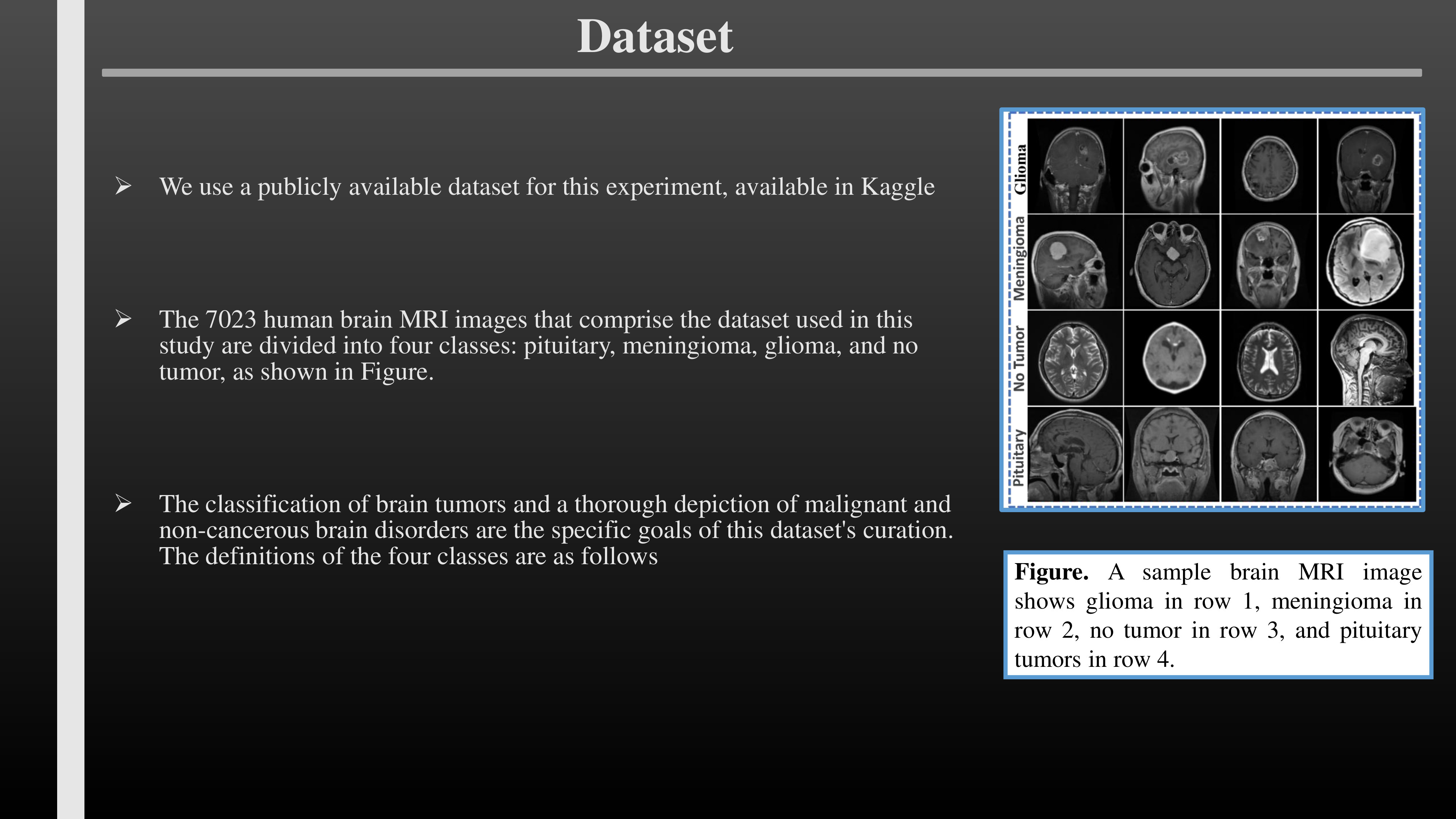 |
We use a publicly available dataset for this experiment, available in Kaggle
The 7023 human brain MRI images that comprise the dataset used in this study are divided into four classes: pituitary, meningioma, glioma, and no tumor, as shown in Figure.
The classification of brain tumors and a thorough depiction of malignant and non-cancerous brain disorders are the specific goals of this dataset's curation. The definitions of the four classes are as follows
|
|
ICACT20250100 Slide.06
[Big slide for presentation]
|
Chrome Text-to-Speach Click!! |
 |
MATERIAL AND METHODS
|
|
ICACT20250100 Slide.05
[Big slide for presentation]
|
Chrome Text-to-Speach Click!! |
 |
To enhance feature extraction, we used a modified ResNet50V2 model that included Squeeze-and-Excitation (SE) architecture.
By recalibrating channel-wise feature responses, SE Module improves CNNs' representational capability by enabling the model to suppress less pertinent features and concentrate on more useful ones adding security
Our updated ResNet50V2 model performed exceptionally well, achieving 99.97% training accuracy and 97.98% validation accuracy. These outcomes show that the proposed method has the potential to be used practically in clinical settings where patient care depends on high-precision
|
|
ICACT20250100 Slide.04
[Big slide for presentation]
|
Chrome Text-to-Speach Click!! |
 |
Brain tumors, whether benign or malignant, are a major issue in neurology because they can cause significant neurological impairment and potentially catastrophic outcomes.
As they grow, this tumor can increase intracranial pressure, which can lead to brain injury, neurological disabilities, and even death.
Early detection and precise classification of brain tumors are essential for effective treatment planning and improved patient outcomes.
For non-invasive tumor diagnosis, magnetic resonance imaging (MRI), which provides fine-grained images of brain anatomy and makes anomaly detection easier, is crucial.
Because manual MRI scan interpretation is time-consuming and prone to human error, automated solutions that assist radiologists in making accurate diagnoses must be developed
|
|
ICACT20250100 Slide.03
[Big slide for presentation]
|
Chrome Text-to-Speach Click!! |
 |
1. INTRODUCTION
|
|
ICACT20250100 Slide.02
[Big slide for presentation]
|
Chrome Text-to-Speach Click!! |
 |
INDEX
Introduction
Methods
Results
Conclusion
|
|
ICACT20250100 Slide.01
[Big slide for presentation]
|
Chrome Text-to-Speach Click!! |
 |
Innovative Deep Learning Strategies for Early Detection of Brain Tumours in MRI Scans with a Modified ResNet50V2 Approach
AUTHOR : RASHADUL ISLAM SUMON, MD ARIFUL ISLAM MAZUMDER , SALMA AKTER, SHAH MUHAMMAD IMTIYAJ UDDIN, HEE-CHEOL KIM
|