ICACT20250002 Slide.01
[Big Slide]
[YouTube] |
Chrome  Click!! |
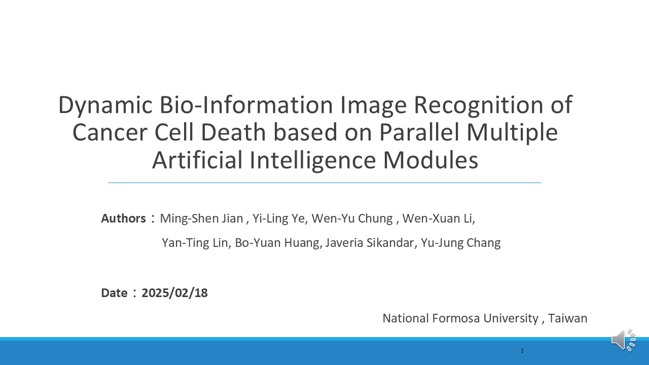 |
Hellow everyone,
It’s an honor to share our research titled "Dynamic Bio-Information Image Recognition of Cancer Cell Death Using Multiple AI Modules." I’m Wen Xuan Lee, from National Formosa University, Taiwan, and I’m excited to explain the main ideas of our study. This project is a team effort to solve key problems in cancer Therapy using advanced AI tools.
|
| ICACT20250002 Slide.02
[Big Slide]
[YouTube] |
Chrome Click!! |
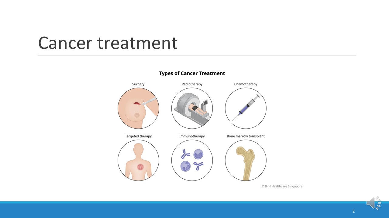 |
Cancer causes over 8.8 million deaths worldwide each year, and the number has been steadily increasing in recent years. Treatment methods for cancer have also been advancing, from traditional surgery and chemotherapy to the current cancer vaccines, targeted therapy, and immunotherapy. Many studies now show that combining targeted therapy with immunotherapy can improve the survival rate of patients.
Among them, immunotherapy has many advantages, such as specificity, memory, and persistence, and it can only attack cancer cells.
So let's talk about immunotherapy.
|
| ICACT20250002 Slide.03
[Big Slide]
[YouTube] |
Chrome Click!! |
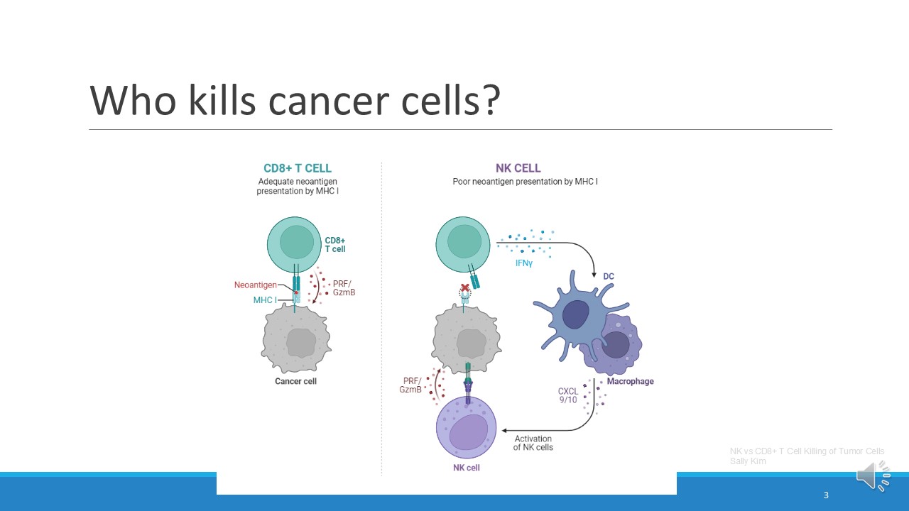 |
Who kills cancer cells? Simply put, there are two types: CD8+ T cells (cytotoxic T cells) and NK cells (natural killer cells).
|
| ICACT20250002 Slide.04
[Big Slide]
[YouTube] |
Chrome Click!! |
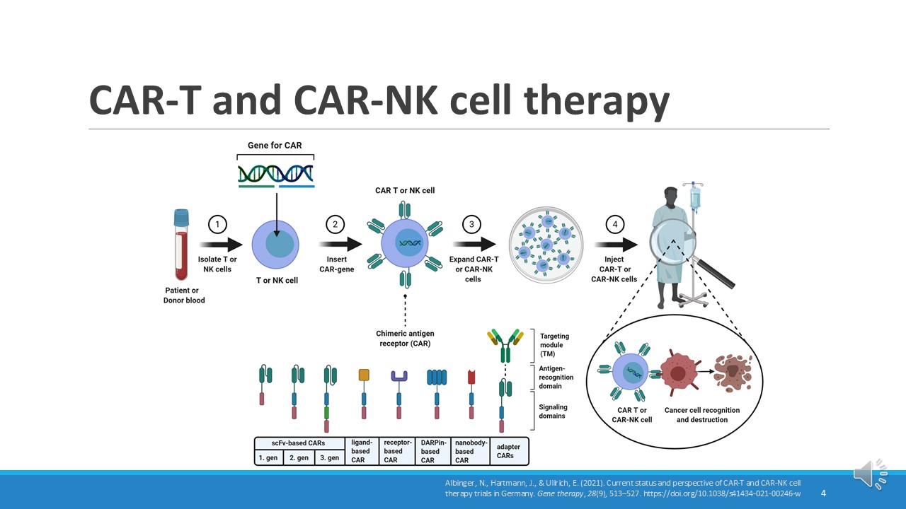 |
So now CART cell or car nk cell therapy has been developed, but its disadvantages are high cost and need to be genetically modified.
|
| ICACT20250002 Slide.05
[Big Slide]
[YouTube] |
Chrome Click!! |
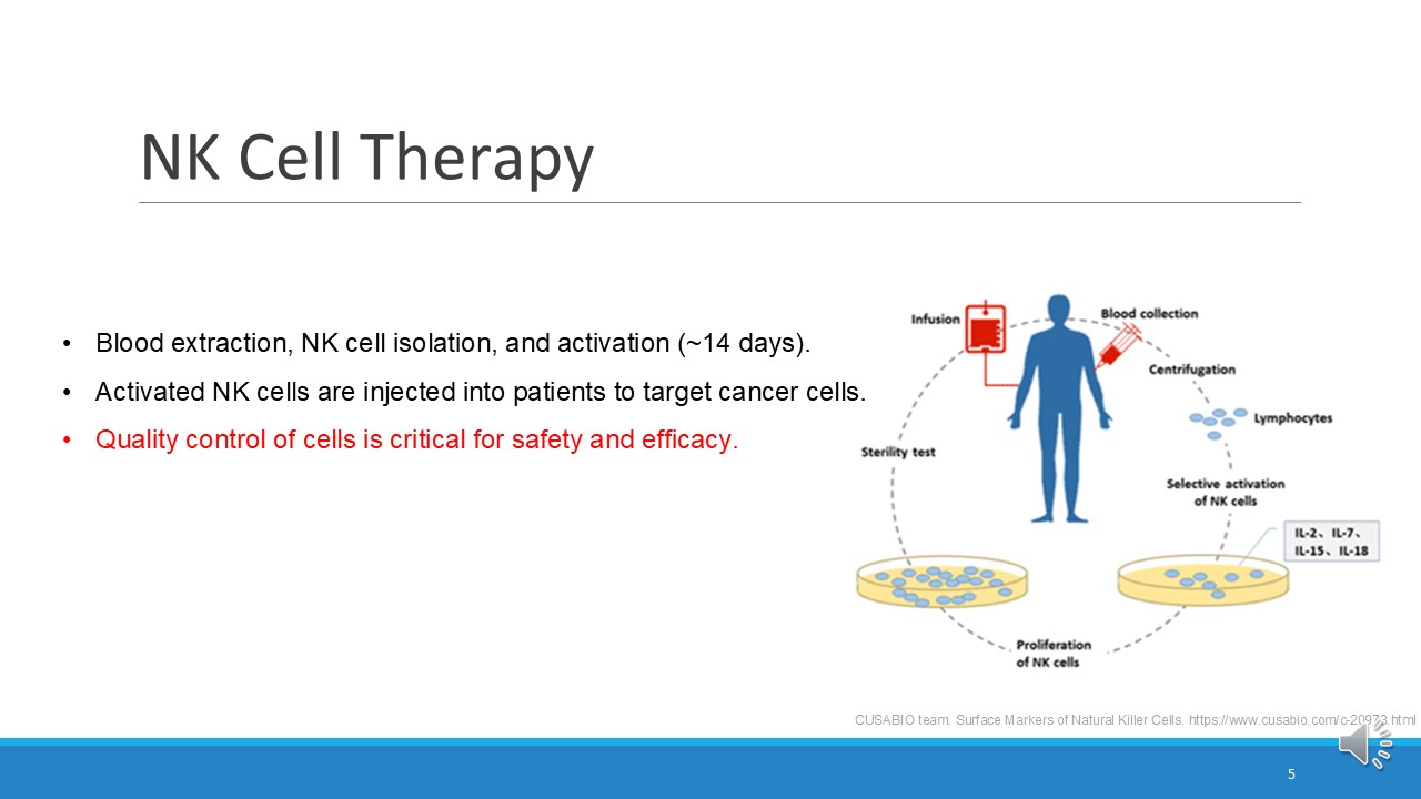 |
So now NK cell immunotherapy has considerable prospects, and it does not require genetic modification.
The NK cell therapy process begins with drawing the patient’s blood, followed by isolating and purifying the NK cells in a laboratory or controlled system. Through cell culture technology and the addition of specific cytokines, NK cells proliferate and activate in large quantities in about 14 days. Finally, these cultured and activated NK cells are reintroduced into the patient to target and destroy cancer cells. Therefore, immune cell therapy attaches great importance to maintaining cell quality and ensuring safety. Whether in immune cell therapy or cytotoxicity experiments, cell survival rate is a key indicator.
|
| ICACT20250002 Slide.06
[Big Slide]
[YouTube] |
Chrome Click!! |
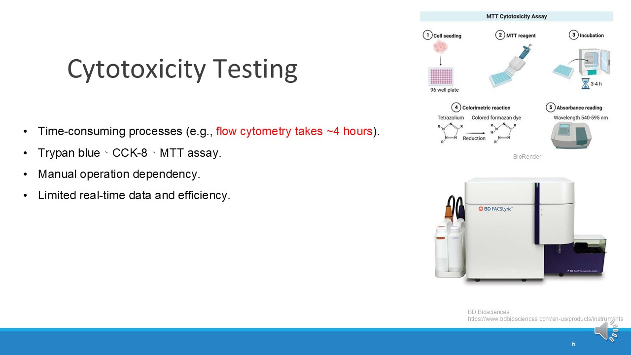 |
In traditional assays for testing cell viability, such as flow cytometry, MTT assay, CCK-8, and trypan blue, a significant amount of time and experimental consumables are required. For example, flow cytometry involves the purchase of antibodies, which are often expensive and come with specificity and time sensitivity.
Moreover, these methods require manual operation.
|
| ICACT20250002 Slide.07
[Big Slide]
[YouTube] |
Chrome Click!! |
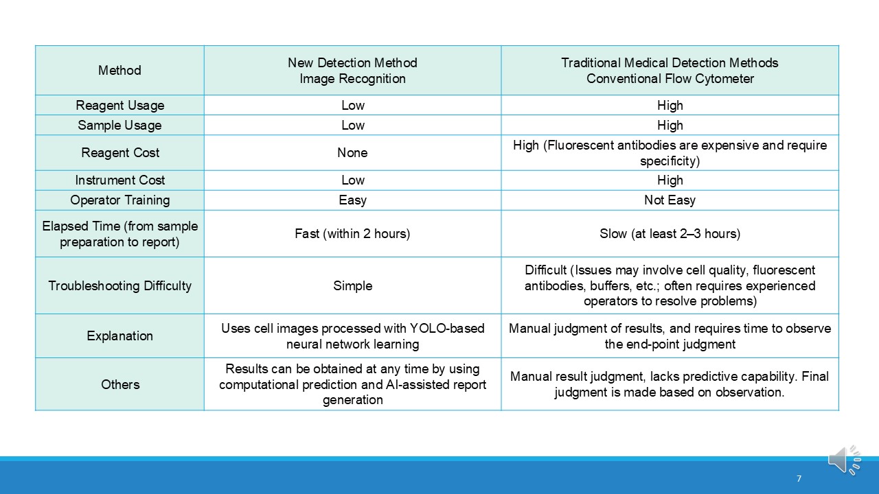 |
Next, let's make a comparison
CCK-8 and MTT take 2-4 hours from sample preparation to results, and MTT is not used for white blood cell detection. The method we developed has the advantages of lower reagent and instrument costs, simpler troubleshooting, faster analysis time (within 2 hours), and automatic prediction and report generation using AI-based technology (YOLO neural network learning). In comparison, traditional flow cytometry requires more resources, higher costs, longer analysis time, and manual judgment, and is therefore more complex and time-consuming.
|
| ICACT20250002 Slide.08
[Big Slide]
[YouTube] |
Chrome Click!! |
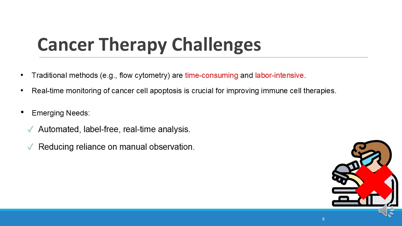 |
Challenges in cancer therapy include the inability to monitor cancer cell death in real time, reliance on manual testing methods, and the use of labels or dyes that increase cost and complexity. These time-consuming methods delay critical results. Faster, automated, and label-free solutions are needed to improve efficiency and enable timely, effective treatments.
|
| ICACT20250002 Slide.09
[Big Slide]
[YouTube] |
Chrome Click!! |
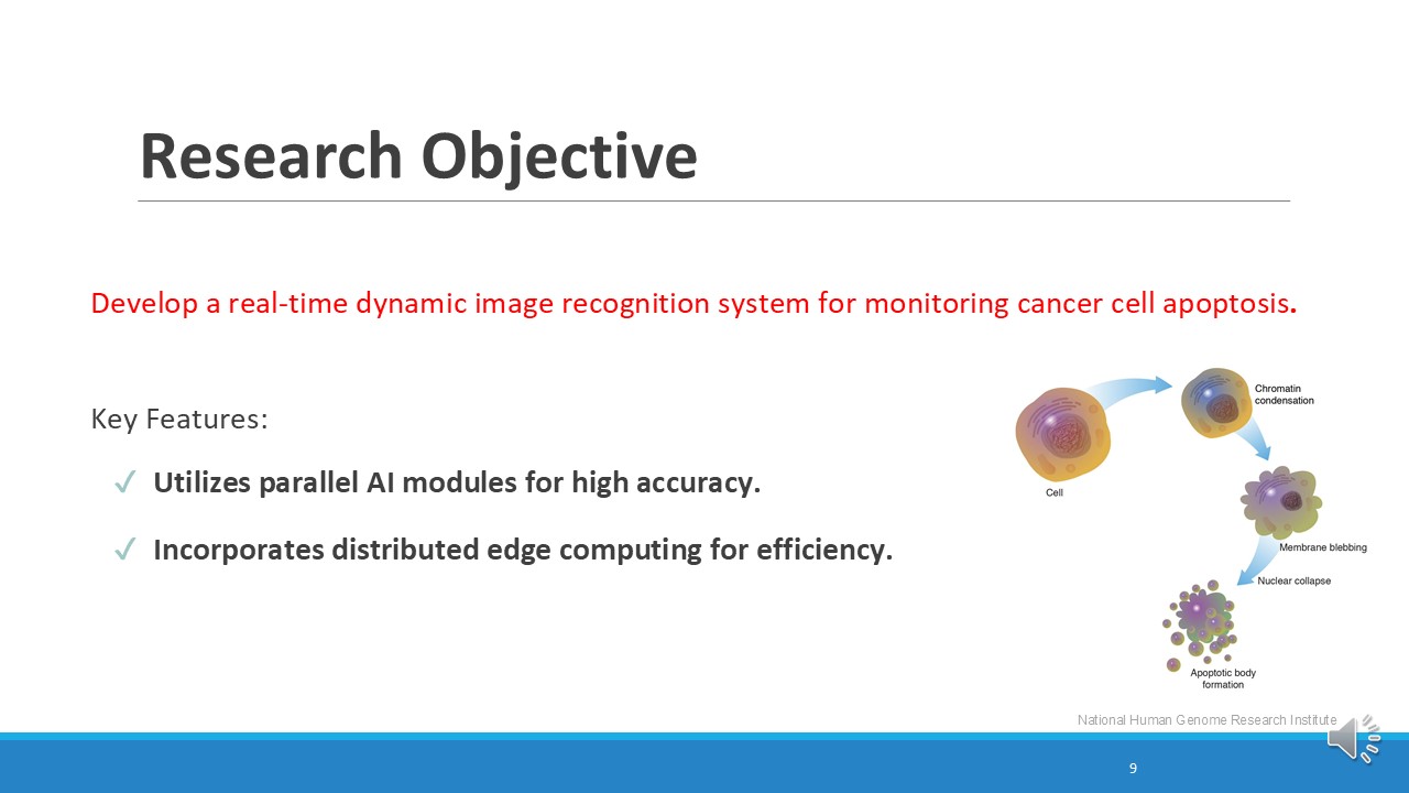 |
Our goal is to develop an advanced system capable of continuously monitoring and tracking cancer cell death in real time. This system would enable researchers and clinicians to observe the dynamic processes of cancer cell apoptosis as they occur, providing critical insights into the immediate effects of therapeutic interventions. By capturing real-time data on cellular changes, such a system would eliminate the delays and limitations associated with traditional methods. Furthermore, it would pave the way for more precise evaluations of treatment efficacy, ultimately contributing to the development of personalized and adaptive cancer therapies that can respond more effectively to patient-specific needs.
|
| ICACT20250002 Slide.10
[Big Slide]
[YouTube] |
Chrome Click!! |
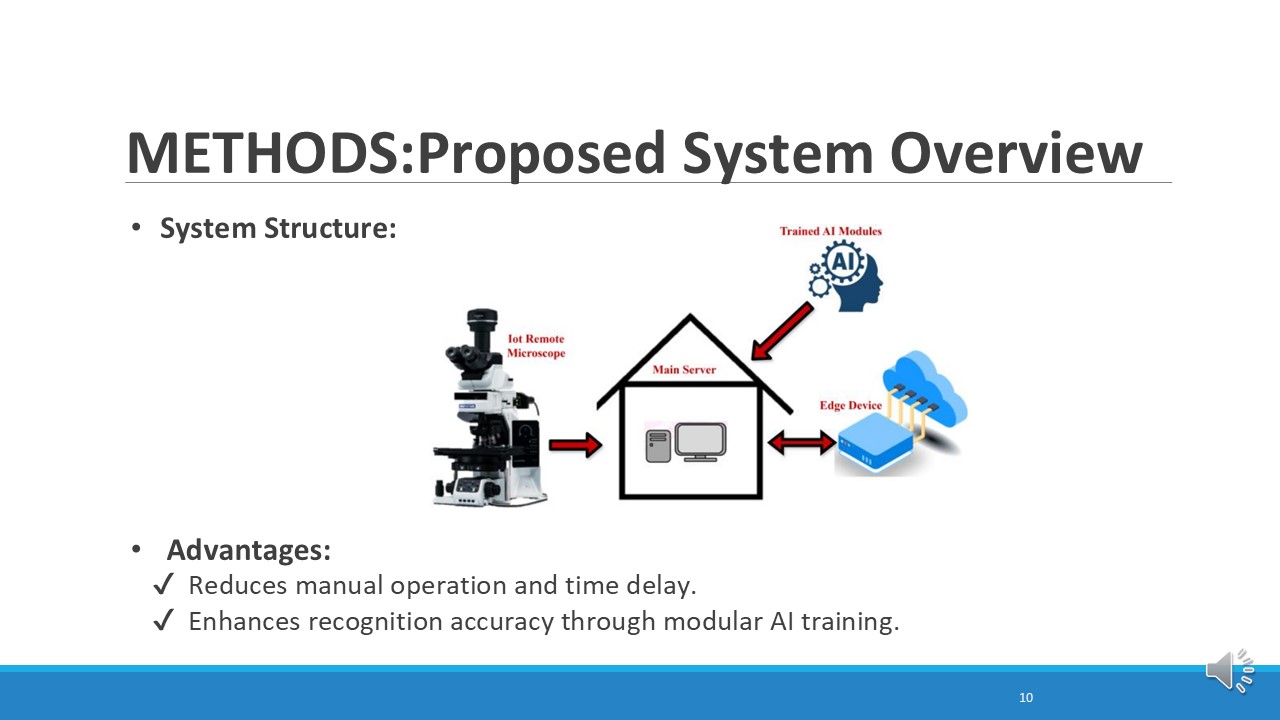 |
This system uses AI and edge computing to speed up and improve data processing. AI analyzes complex data quickly, while edge computing processes it locally, reducing delays and server dependency. This automation frees up researchers for critical tasks, streamlines workflows, and ensures precise, efficient results.
|
| ICACT20250002 Slide.11
[Big Slide]
[YouTube] |
Chrome Click!! |
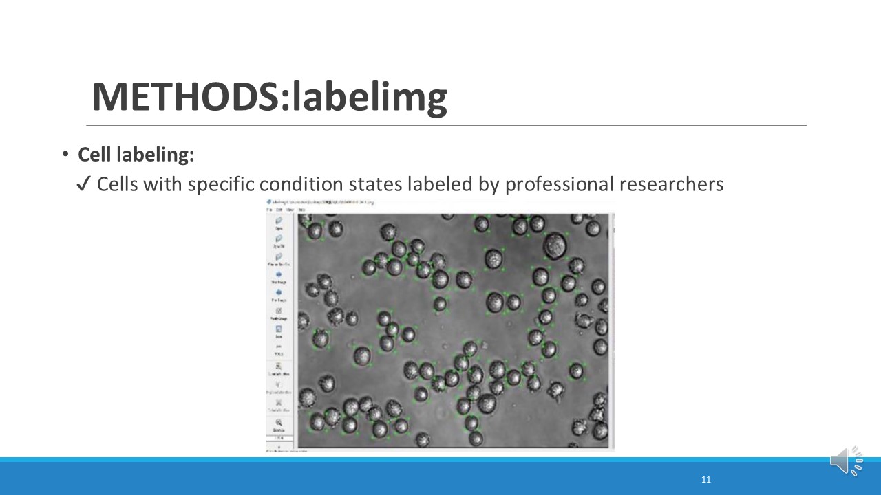 |
In the cell labeling process,
professional researchers use software called LabelImg to annotate cells with specific condition states.
The labeled images are then exported to the training dataset,
which is used for YOLO model training.
|
| ICACT20250002 Slide.12
[Big Slide]
[YouTube] |
Chrome Click!! |
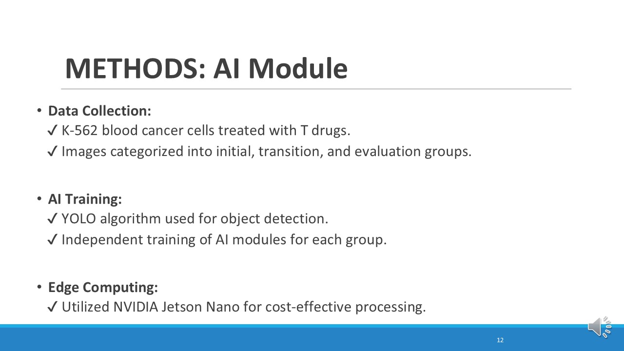 |
We collected images of K-562 blood cancer cells treated with a drug.
The images were grouped into three stages: beginning, changing, and final.
We used the YOLO to train the AI to detect and classify the images.
Edge Computing
The NVIDIA Jetson Nano was used for fast and cost-effective processing at the edge.
|
| ICACT20250002 Slide.13
[Big Slide]
[YouTube] |
Chrome Click!! |
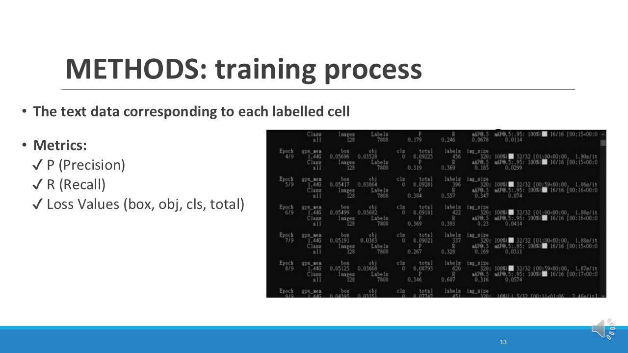 |
Here, we can see the training process and key metrics from Epoch 4 to Epoch 9.
we have the Precision (P) and Recall (R).
Precision measures how many of the detected cells are correct,
while recall shows how many of the actual cells are successfully detected
And
we have the loss values,
These numbers help us understand how the model is improving during training.
|
| ICACT20250002 Slide.14
[Big Slide]
[YouTube] |
Chrome Click!! |
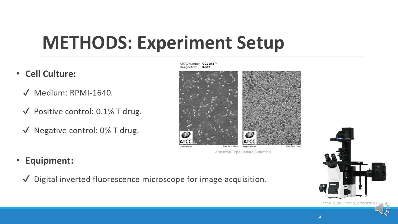 |
Cancer cells were grown in RPMI-1640 medium.
Positive controls were treated with 0.1% of the drug, while negative controls without drugs.
Images were captured using a digital fluorescence microscope.
|
| ICACT20250002 Slide.15
[Big Slide]
[YouTube] |
Chrome Click!! |
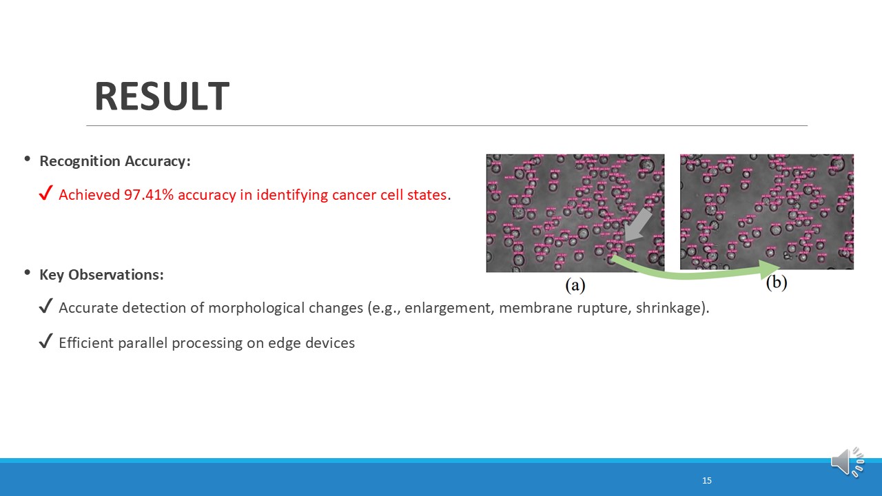 |
Our system achieved 97.41% accuracy in identifying and classifying cancer cell states, proving its precision and reliability. It detects key changes like cell swelling, membrane breaking, and shrinking, offering valuable insights into cancer progression and treatment effects.
|
| ICACT20250002 Slide.16
[Big Slide]
[YouTube] |
Chrome Click!! |
 |
Video Presentation of Verification |
| ICACT20250002 Slide.17
[Big Slide]
[YouTube] |
Chrome Click!! |
 |
This system revolutionizes cancer research and treatment by using AI and edge computing to automate real-time testing, even in remote hospitals. It improves accessibility, reduces lab waste, supports sustainability, and lowers costs, making healthcare more efficient and eco-friendly.
|
| ICACT20250002 Slide.18
[Big Slide]
[YouTube] |
Chrome Click!! |
 |
This capability not only enhances the understanding of cellular responses to treatments but also supports researchers and clinicians in making more informed decisions regarding therapeutic strategies. The system's high accuracy and detailed recognition ability highlight its potential as a powerful tool in advancing cancer research and improving patient outcomes.
|
| ICACT20250002 Slide.19
[Big Slide]
[YouTube] |
Chrome Click!! |
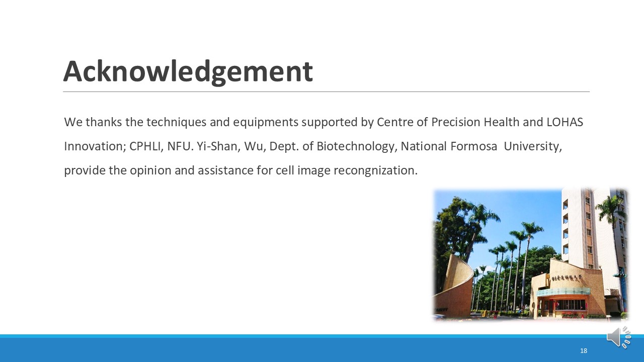 |
We thank the Centre of Precision Health and LOHAS Innovation at National Formosa University for their support, and Yi-Shan Wu for her help with cell image recognition.
|
| ICACT20250002 Slide.20
[Big Slide]
[YouTube] |
Chrome Click!! |
 |
Thank you |