ICACT20260052 Slide.01
[Big Slide] |
Chrome  Click!! |
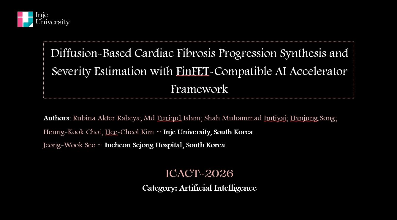 |
Hello everyone. This is Rubina Akter Rabeya, a Ph.D. student in Artificial Intelligence from Inje University, South Korea. In this presentation, I will discuss our research on diffusion-based cardiac ischemic fibrosis progression synthesis and severity estimation using histopathology images. The study also introduces a FinFET-compatible AI accelerator framework, aiming to bridge advanced generative AI with future low-power and edge-based clinical deployment. This work lies at the intersection of medical image analysis, generative AI, and nanoelectronics. |
| ICACT20260052 Slide.02
[Big Slide]
|
Chrome Click!! |
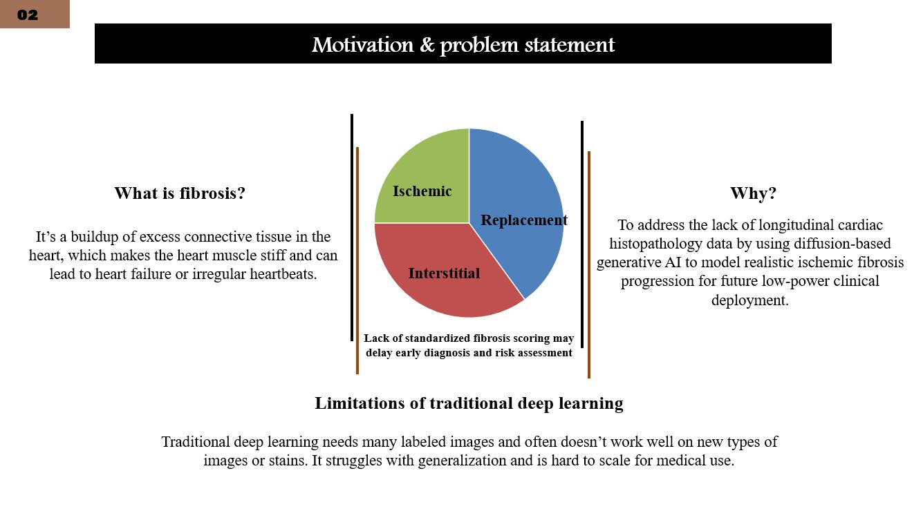 |
Cardiac ischemic fibrosis is a major pathological outcome of aging and myocardial infarction, significantly impacting cardiac function and patient prognosis. Histopathology remains the gold standard for fibrosis assessment, but acquiring longitudinal tissue samples over time is clinically impractical. As a result, understanding fibrosis progression dynamics is extremely challenging. This motivates the use of generative AI models to simulate disease progression from static histological data. |
| ICACT20260052 Slide.03
[Big Slide]
|
Chrome Click!! |
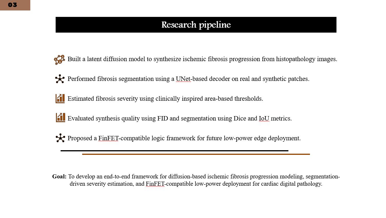 |
The overall goal of this research is to develop a unified framework that can model fibrosis progression, accurately segment ischemic regions, estimate disease severity, and evaluate feasibility for low-power deployment. As shown in this pipeline, we integrate diffusion-based synthesis, segmentation-based analysis, and hardware-aware design into a single end-to-end system. |
| ICACT20260052 Slide.04
[Big Slide]
|
Chrome Click!! |
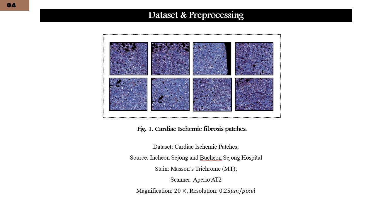 |
This slide presents examples of ischemic fibrosis patches extracted from cardiac histopathology whole-slide images. We used 5,000 Masson’s trichrome-stained patches, each of size 512 by 512 pixels, manually annotated by expert pathologists. The dataset focuses specifically on ischemic fibrosis regions, ensuring clinical relevance and annotation reliability. |
| ICACT20260052 Slide.05
[Big Slide]
|
Chrome Click!! |
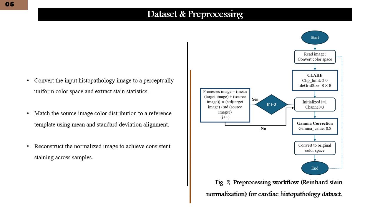 |
Histopathology images often suffer from staining variability and noise. To address this, we applied a standardized preprocessing pipeline. This includes noise reduction, contrast enhancement, and Reinhard stain normalization, which aligns the color distribution of all images to a reference template. These steps improve visual consistency and enhance model generalization. |
| ICACT20260052 Slide.06
[Big Slide]
|
Chrome Click!! |
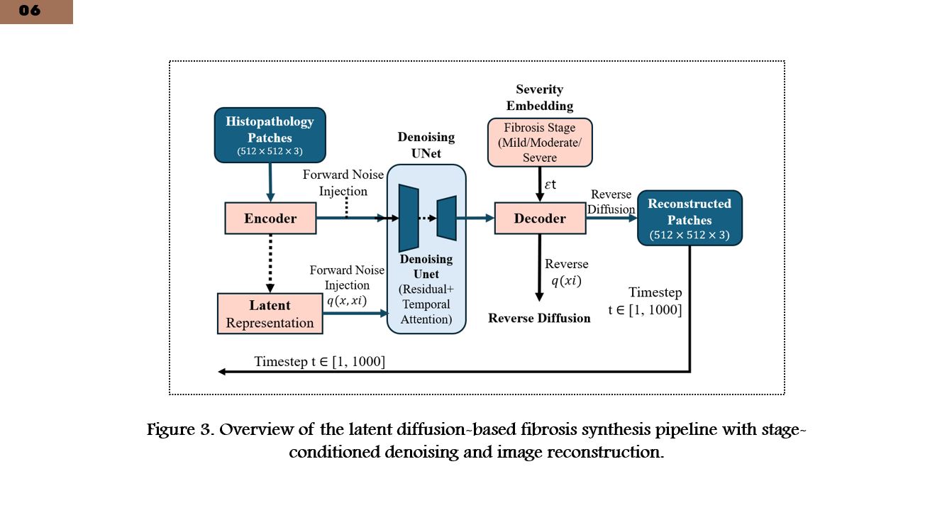 |
This figure illustrates the overall diffusion-based synthesis workflow. Input images are first encoded into a latent representation. Gaussian noise is then iteratively added and removed through a controlled diffusion process. Finally, the denoised latent representation is decoded to generate realistic fibrosis images conditioned on different disease severity stages. |
| ICACT20260052 Slide.07
[Big Slide]
|
Chrome Click!! |
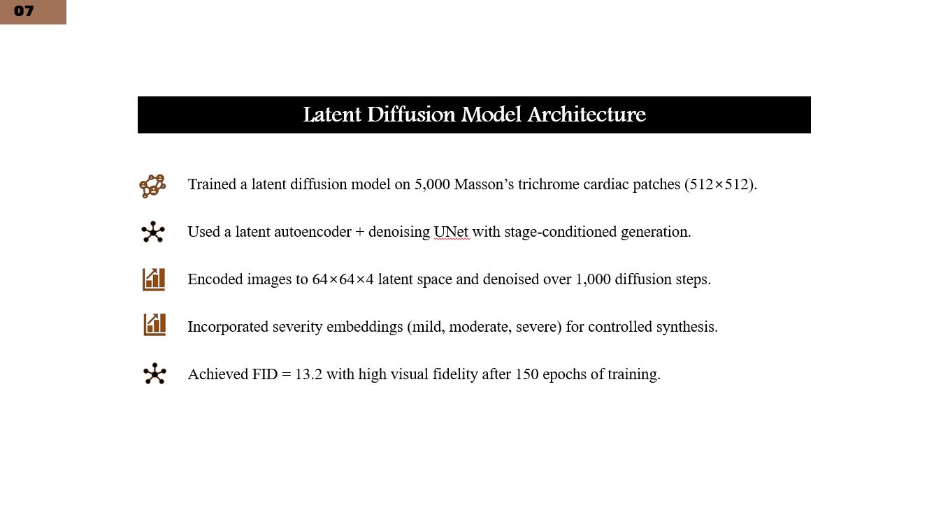 |
Here, we present the architecture of the latent diffusion model. A convolutional autoencoder compresses images into a low-dimensional latent space. A UNet-based denoising network operates in this latent space, progressively removing noise. Severity stage embeddings are injected into the network, enabling controlled synthesis of mild, moderate, and severe fibrosis patterns. |
| ICACT20260052 Slide.08
[Big Slide]
|
Chrome Click!! |
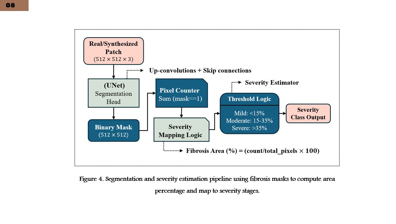 |
After synthesis, both real and generated images are passed through a segmentation pipeline. A UNet-based model identifies fibrosis regions at the pixel level. The resulting segmentation masks are then used to compute fibrosis area percentages, which form the basis for severity estimation. |
| ICACT20260052 Slide.09
[Big Slide]
|
Chrome Click!! |
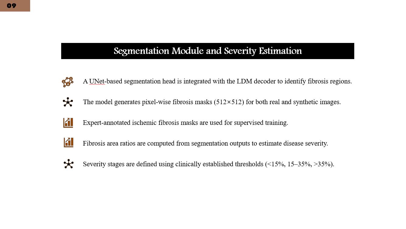 |
This slide focuses on segmentation and severity estimation. The segmentation model accurately localizes ischemic fibrosis regions. Severity is estimated using clinically inspired area-based thresholds, categorizing fibrosis into mild, moderate, or severe stages. This approach provides an interpretable and clinically meaningful severity assessment. |
| ICACT20260052 Slide.10
[Big Slide]
|
Chrome Click!! |
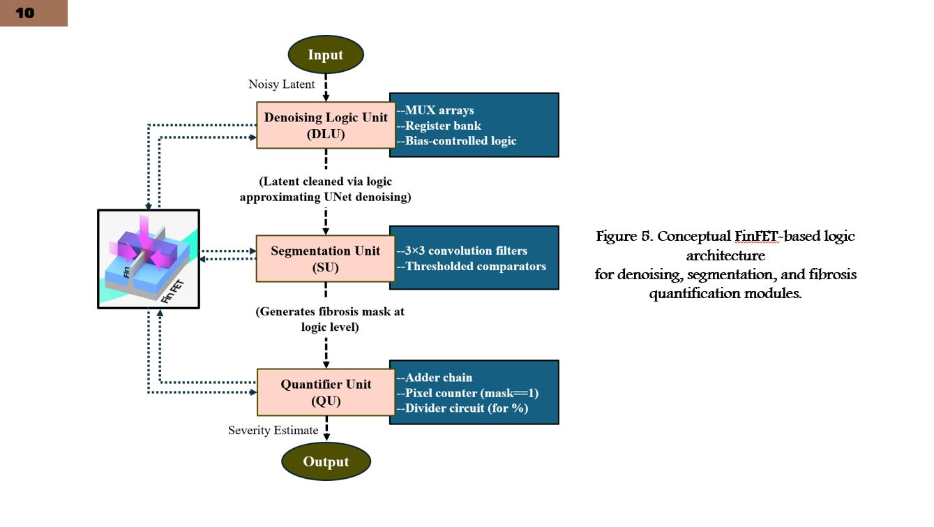 |
To enable future edge deployment, we propose a conceptual FinFET-compatible architecture. The AI pipeline is mapped into three main logic modules: denoising, segmentation, and quantification. This design emphasizes low power consumption and hardware efficiency, suitable for medical edge devices. |
| ICACT20260052 Slide.11
[Big Slide]
|
Chrome Click!! |
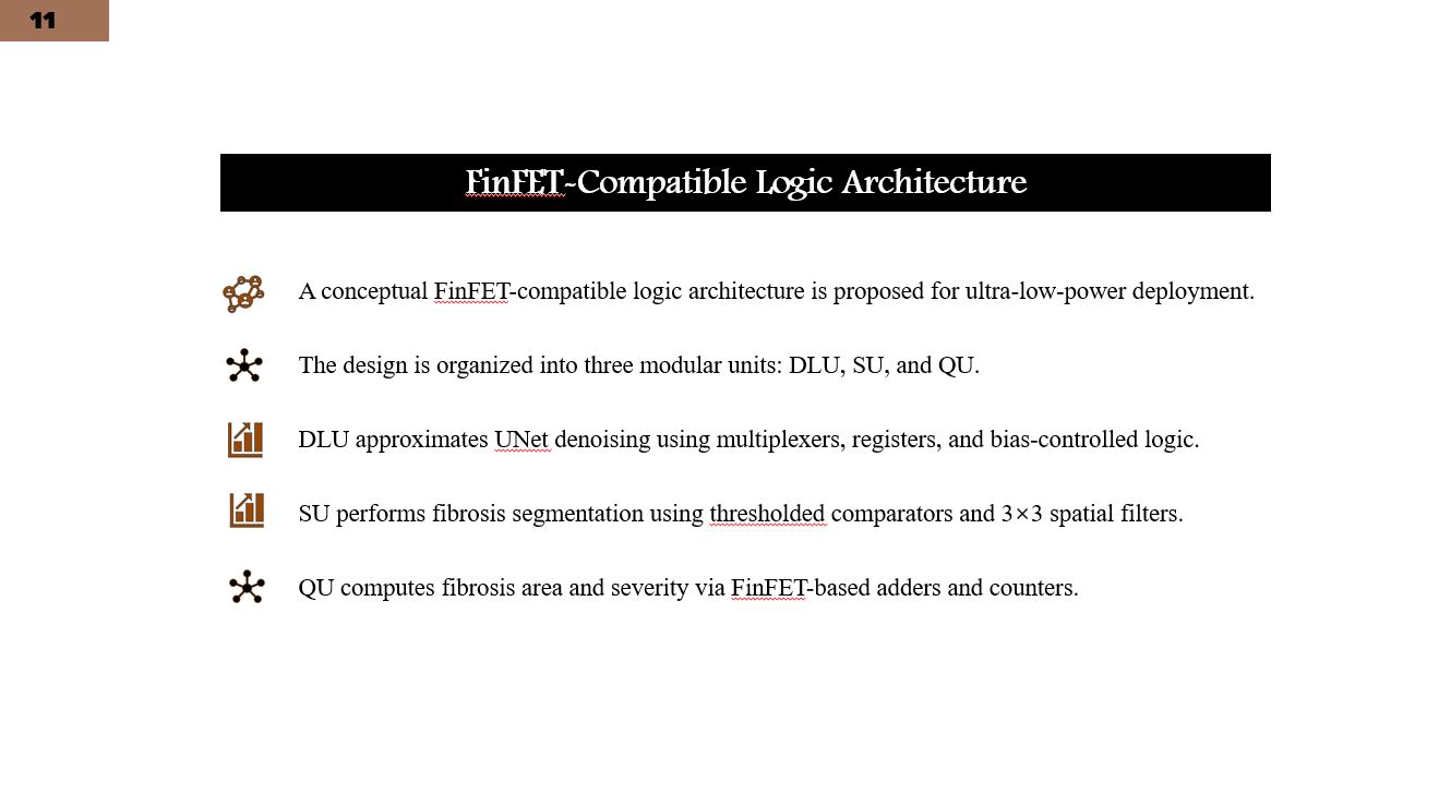 |
This slide details the internal logic design of the FinFET-based modules. Denoising is approximated using multiplexers, registers, and bias-controlled logic. Segmentation relies on spatial filtering and comparator circuits, while quantification uses adders and counters to compute fibrosis area percentages. |
| ICACT20260052 Slide.12
[Big Slide]
|
Chrome Click!! |
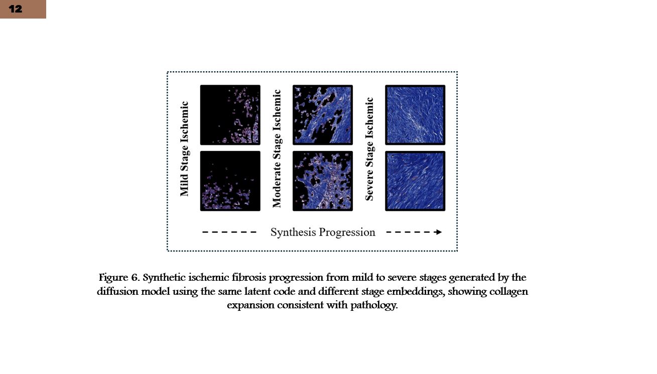 |
Here, we show synthetic ischemic fibrosis progression from mild to severe stages. All images are generated using the same latent code with different stage embeddings. The visual results demonstrate biologically plausible expansion of fibrotic regions, highlighting the model’s ability to capture disease progression patterns. |
| ICACT20260052 Slide.13
[Big Slide]
|
Chrome Click!! |
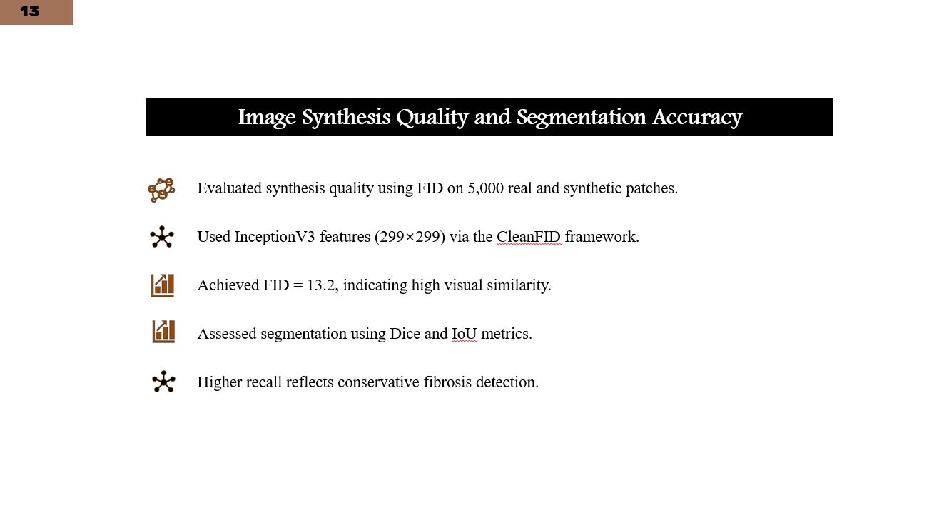 |
Synthesis quality was evaluated using the Frechet Inception Distance, achieving a score of 13.2, which indicates strong similarity between real and generated images. Segmentation performance was evaluated using Dice and IoU metrics, achieving a Dice score of 0.91, demonstrating accurate fibrosis localization. |
| ICACT20260052 Slide.14
[Big Slide]
|
Chrome Click!! |
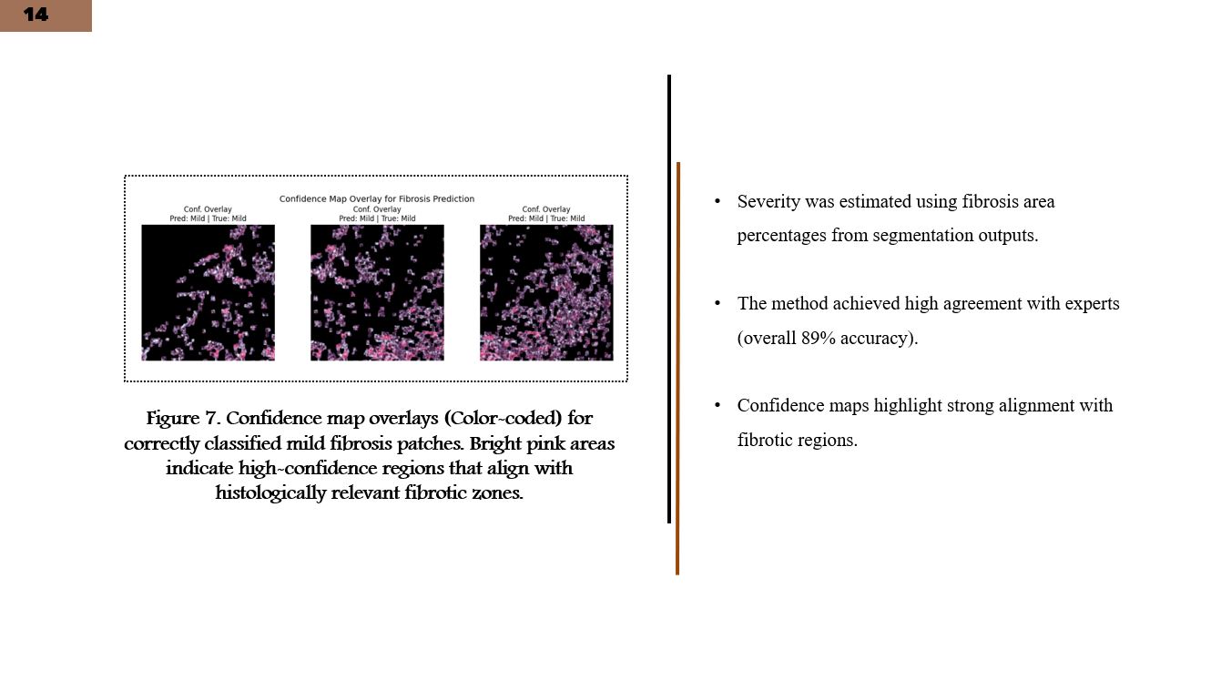 |
This slide presents confidence visualization maps highlighting regions strongly associated with fibrosis severity. Severity estimation achieved approximately 89 percent accuracy when compared with expert annotations, confirming strong alignment with clinical grading. |
| ICACT20260052 Slide.15
[Big Slide]
|
Chrome Click!! |
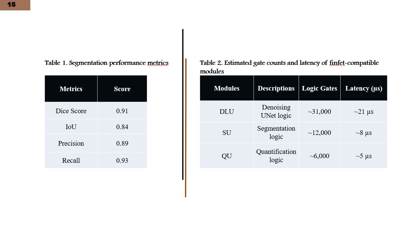 |
This slide summarizes quantitative performance metrics, including segmentation accuracy, severity estimation results, and hardware-related metrics such as gate counts and latency. These results demonstrate consistency between AI performance and hardware feasibility. |
| ICACT20260052 Slide.16
[Big Slide]
|
Chrome Click!! |
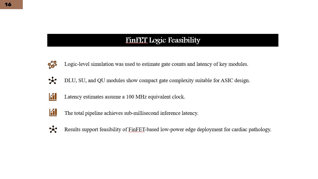 |
Logic-level simulations were conducted assuming a 100 megahertz equivalent clock. The results show compact gate complexity and sub-millisecond latency across modules, supporting feasibility for future low-power ASIC-based deployment in cardiac digital pathology. |
| ICACT20260052 Slide.17
[Big Slide]
|
Chrome Click!! |
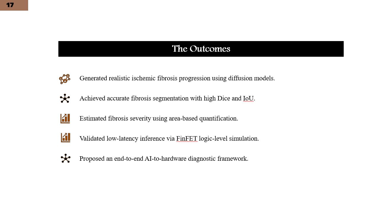 |
Overall, this research demonstrates a unified framework that successfully integrates diffusion-based fibrosis synthesis, accurate segmentation, clinically interpretable severity estimation, and hardware-aware feasibility analysis. |
| ICACT20260052 Slide.18
[Big Slide]
|
Chrome Click!! |
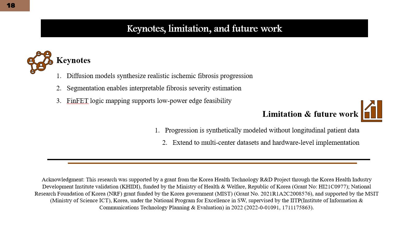 |
Key contributions include realistic disease progression modeling and a hardware-aware design perspective. A key limitation is reliance on synthetic progression rather than real longitudinal data. Future work will focus on multi-center validation and hardware-level prototyping. |
| ICACT20260052 Slide.19
[Big Slide]
|
Chrome Click!! |
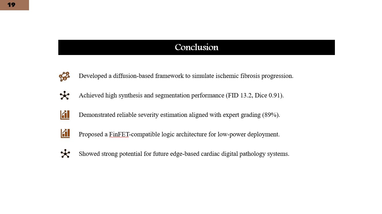 |
In conclusion, this work highlights the potential of diffusion-based generative AI combined with FinFET-compatible design to enable future edge-based, low-power cardiac diagnostic systems, particularly for aging-related cardiovascular diseases. |
| ICACT20260052 Slide.20
[Big Slide]
|
Chrome Click!! |
 |
Thank you for your attention. Please ask me if you have any questions. |