ICACT20260266 Slide.01
[Big Slide] |
Chrome  Click!! |
 |
Good morning/afternoon, everyone.
My name is Kiramat Ullah, and today I am presenting our work titled “CardioViT-CAM: Self-Supervised Vision Transformer with Class Activation Mapping for Interpretable Congenital Heart Disease Detection.” This work is conducted in collaboration with my co-authors. In this research, we propose an explainable deep learning framework for detecting congenital heart defects using echocardiography data. |
| ICACT20260266 Slide.02
[Big Slide]
|
Chrome Click!! |
 |
Here is the main content of my presentation. |
| ICACT20260266 Slide.03
[Big Slide]
|
Chrome Click!! |
 |
Congenital Heart Disease, or CHD, refers to structural abnormalities of the heart that are present at birth.
It is a major global health problem affecting millions of newborns every year.
The global prevalence ranges from approximately 7 to 12 cases per 1000 live births, and CHD remains a leading cause of mortality in children.
In Pakistan alone, around 60,000 babies are born with CHD annually, highlighting the need for early diagnosis and treatment. |
| ICACT20260266 Slide.04
[Big Slide]
|
Chrome Click!! |
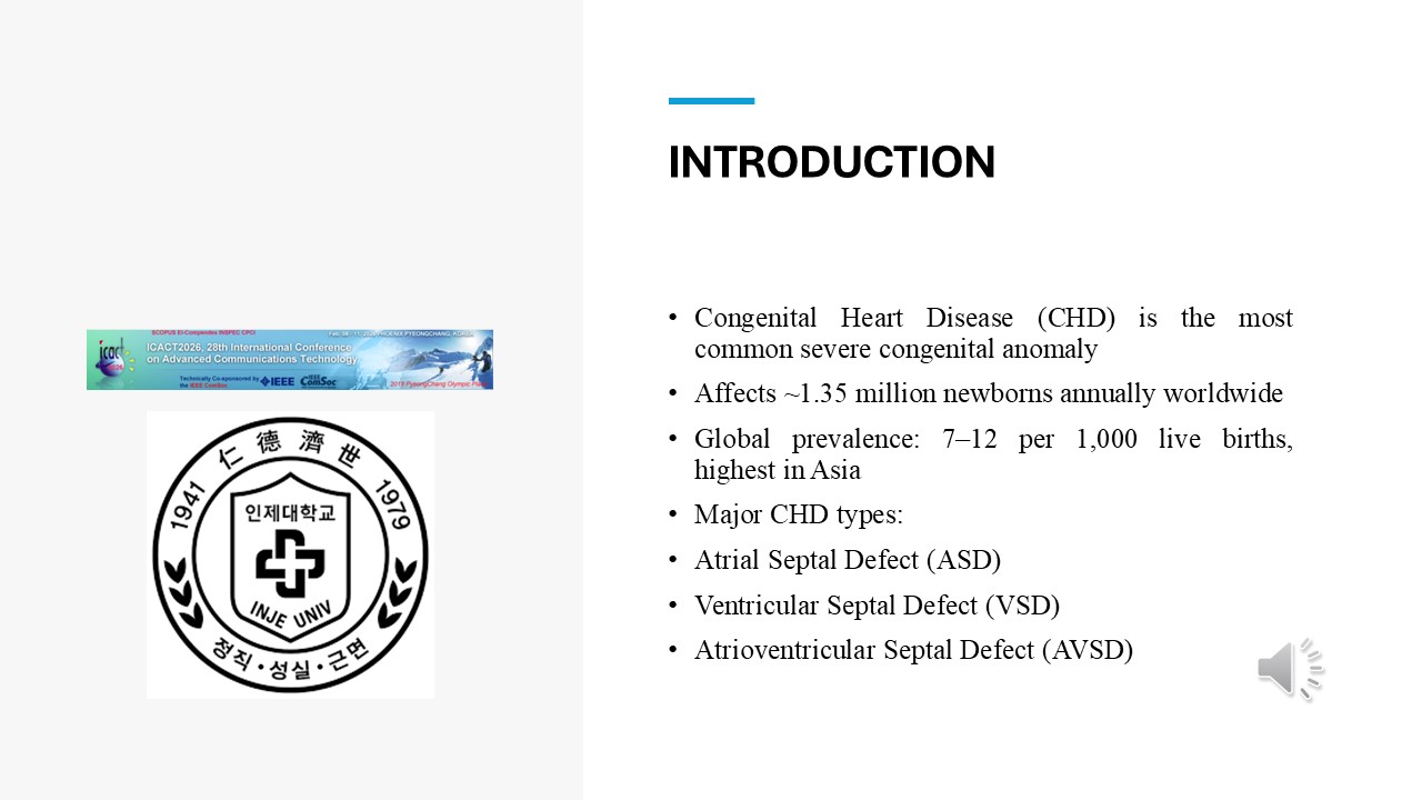 |
Among different congenital heart defects, this study focuses on three major types:
Atrial Septal Defect (ASD), which is a hole between the upper chambers of the heart.
Ventricular Septal Defect (VSD), which occurs between the lower chambers.
Atrioventricular Septal Defect (AVSD), which is a more complex defect involving both upper and lower chambers.
These defects significantly affect blood flow and cardiac function. |
| ICACT20260266 Slide.05
[Big Slide]
|
Chrome Click!! |
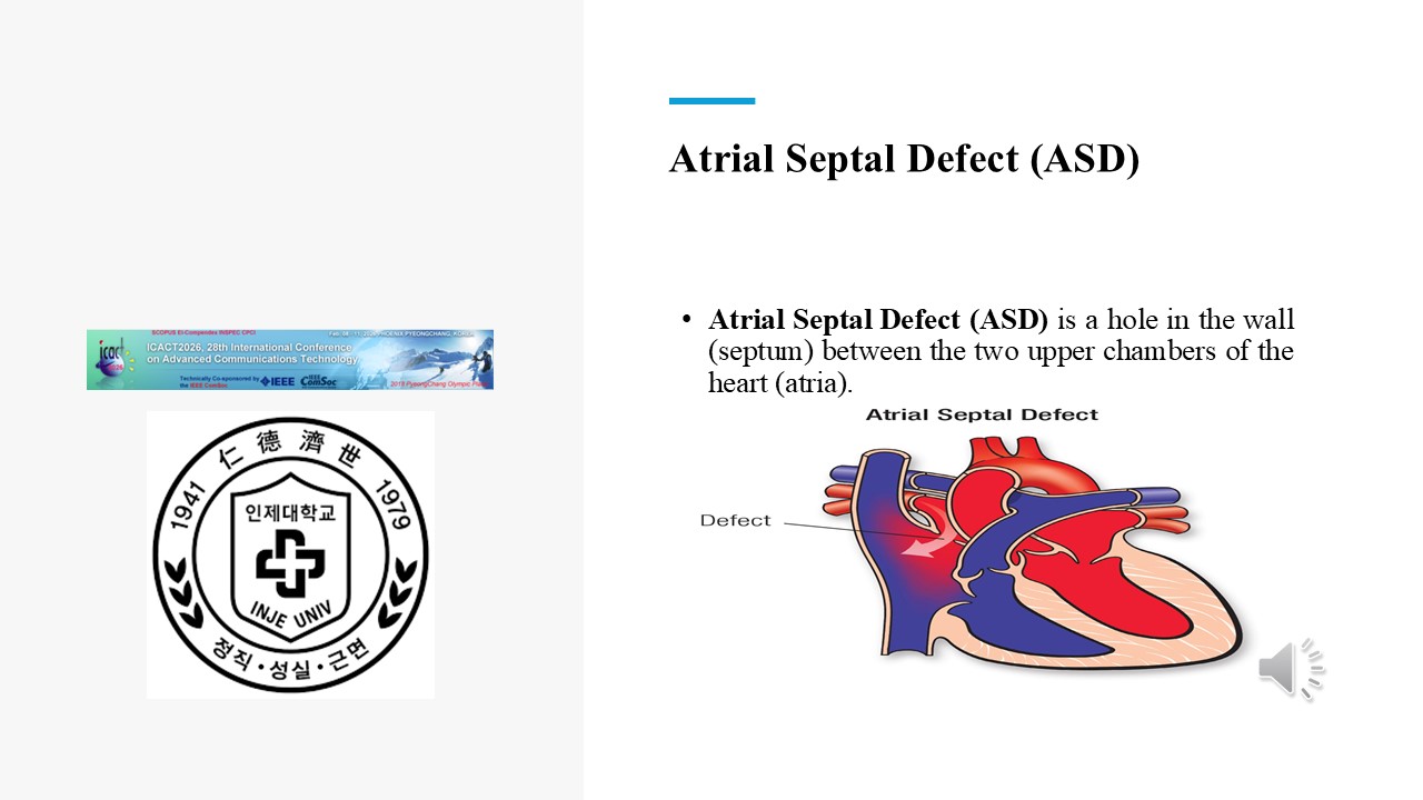 |
Atrial Septal Defect (ASD) is a hole in the wall (septum) between the two upper chambers of the heart (atria).
|
| ICACT20260266 Slide.06
[Big Slide]
|
Chrome Click!! |
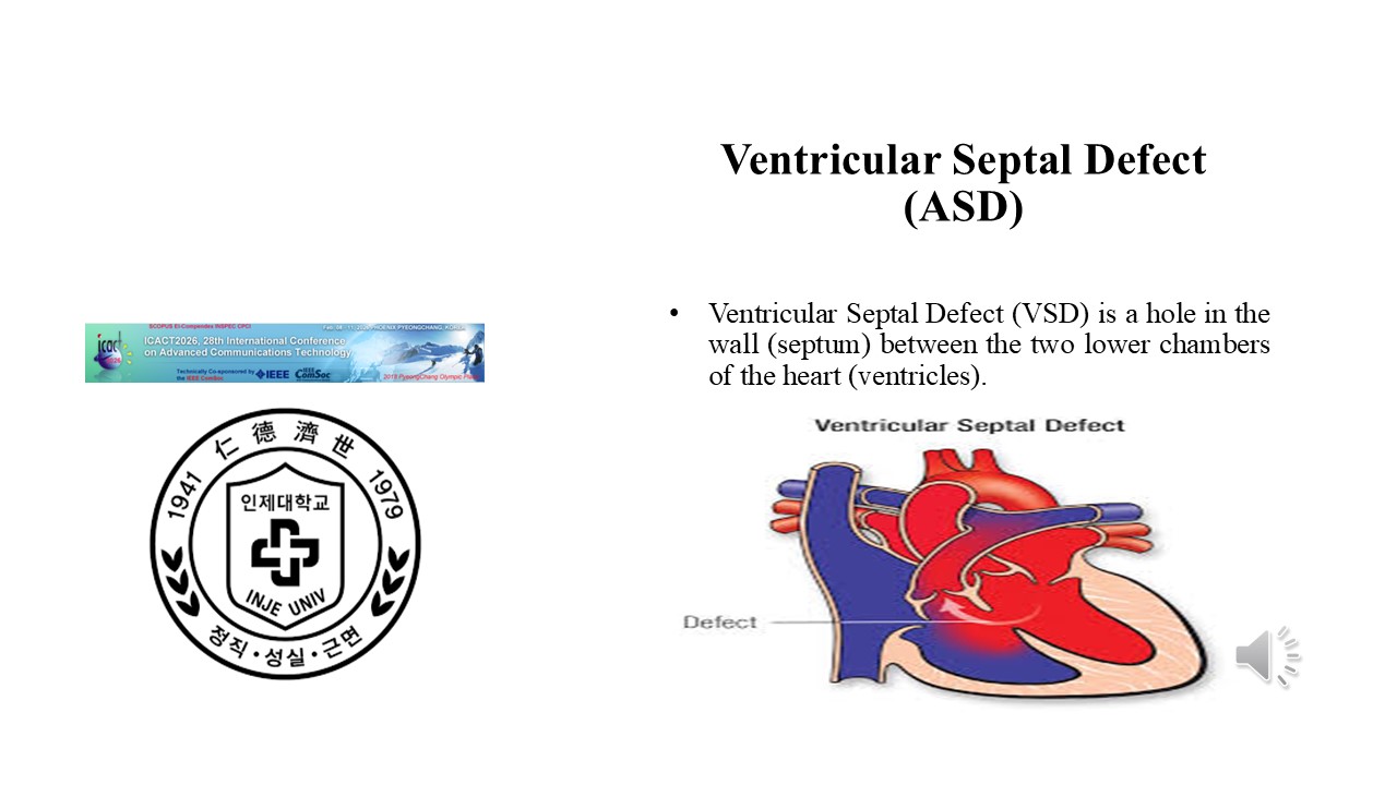 |
Ventricular Septal Defect (VSD) is a hole in the wall (septum) between the two lower chambers of the heart (ventricles).
|
| ICACT20260266 Slide.07
[Big Slide]
|
Chrome Click!! |
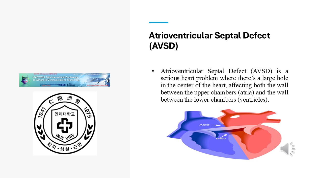 |
Atrioventricular Septal Defect (AVSD) is a serious heart problem where there’s a large hole in the center of the heart, affecting both the wall between the upper chambers (atria) and the wall between the lower chambers (ventricles).
|
| ICACT20260266 Slide.08
[Big Slide]
|
Chrome Click!! |
 |
Transthoracic echocardiography, including 2D imaging and Color Doppler, is the primary diagnostic tool for CHD. It provides visualization of cardiac structure and blood flow patterns.
However, early screening of ASD, VSD, and AVSD remains challenging, even for experienced cardiologists. |
| ICACT20260266 Slide.09
[Big Slide]
|
Chrome Click!! |
 |
Current CHD diagnosis is highly dependent on expert interpretation, which can be time-consuming and subjective. This may lead to missed or delayed diagnosis. Therefore, there is a strong need to develop an accurate and interpretable AI-based system to support clinicians in early CHD detection. |
| ICACT20260266 Slide.10
[Big Slide]
|
Chrome Click!! |
 |
For this study, we used a private echocardiography dataset collected from Lady Reading Hospital, Pakistan, between 2022 and 2024.The dataset contains: 1600 TTE videos Approximately 74,000 extracted frames From these, 6000 images were labeled by two senior cardiologists for supervised training.
All data were anonymized and approved by the Institutional Review Board. |
| ICACT20260266 Slide.11
[Big Slide]
|
Chrome Click!! |
 |
Our proposed framework, called CardioViT-CAM, consists of two main stages.First, we perform self-supervised pretraining using a Masked Autoencoder with a Vision Transformer to learn general cardiac features. Second, we perform supervised fine-tuning for classification of ASD, VSD, and AVSD.
Additionally, we apply Grad-CAM++ to provide interpretable visualization of model predictions. |
| ICACT20260266 Slide.12
[Big Slide]
|
Chrome Click!! |
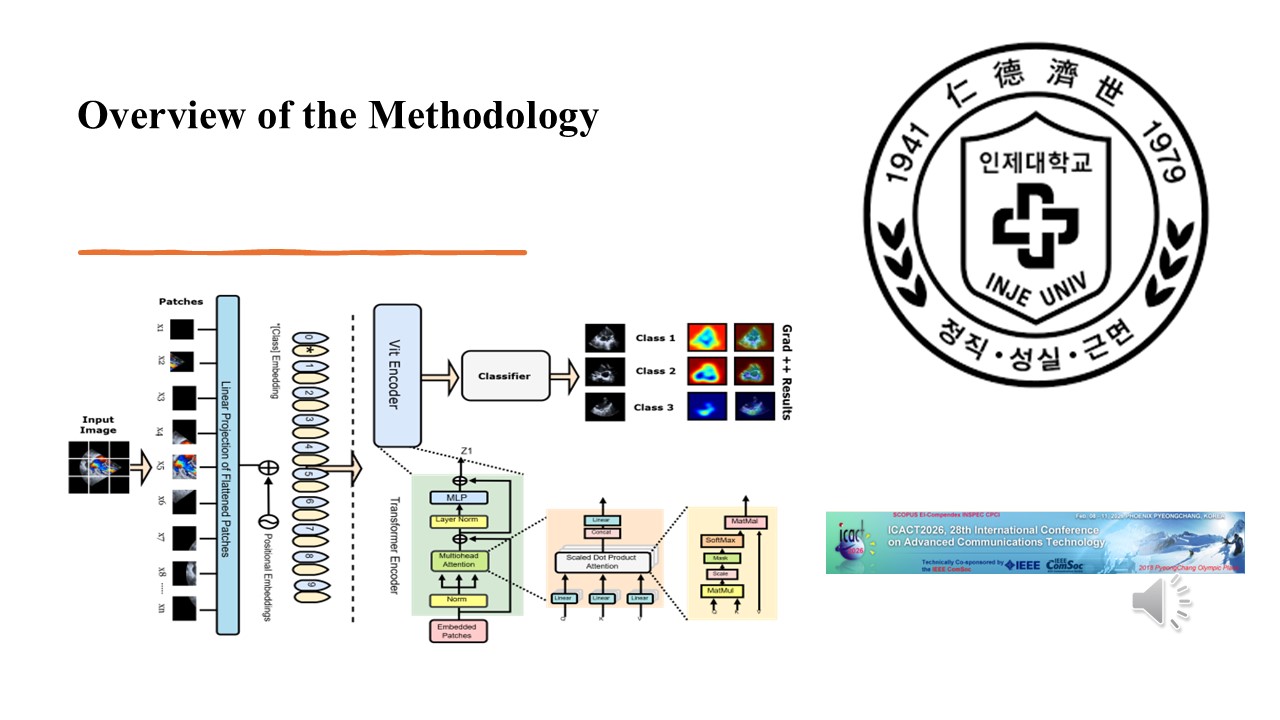 |
This figure illustrates the overall architecture of the proposed system. Unlabeled echocardiography frames are first used for MAE pretraining. Then, the pretrained encoder is fine-tuned using labeled data for classification. Finally, Grad-CAM++ highlights diagnostic regions that contribute to model predictions. |
| ICACT20260266 Slide.13
[Big Slide]
|
Chrome Click!! |
 |
We evaluated multiple visualization methods including Grad-CAM, Score-CAM, and Grad-CAM++. All models used the same pretrained architecture. The only difference was the visualization technique. Among them, Grad-CAM++ achieved the best performance across all evaluation metrics |
| ICACT20260266 Slide.14
[Big Slide]
|
Chrome Click!! |
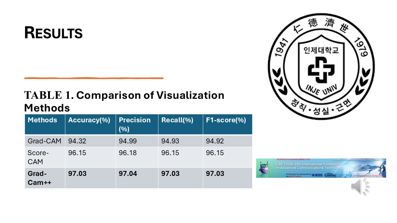 |
This table summarizes the classification performance.
Grad-CAM++ achieved:
97.03% Accuracy
97.04% Precision
97.03% Recall
97.03% F1 Score
These results demonstrate strong diagnostic capability of the proposed framework. |
| ICACT20260266 Slide.15
[Big Slide]
|
Chrome Click!! |
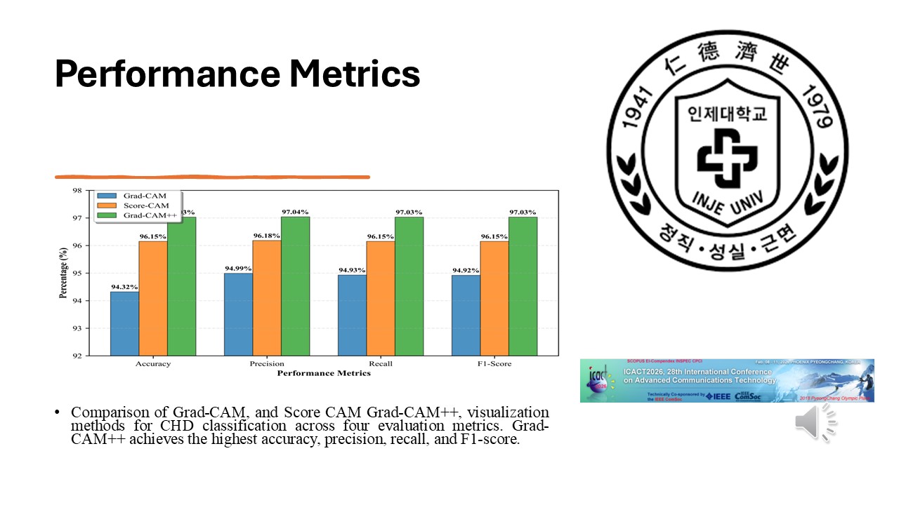 |
Comparison of Grad-CAM, and Score CAM Grad-CAM++, visualization methods for CHD classification across four evaluation metrics. Grad-CAM++ achieves the highest accuracy, precision, recall, and F1-score.
|
| ICACT20260266 Slide.16
[Big Slide]
|
Chrome Click!! |
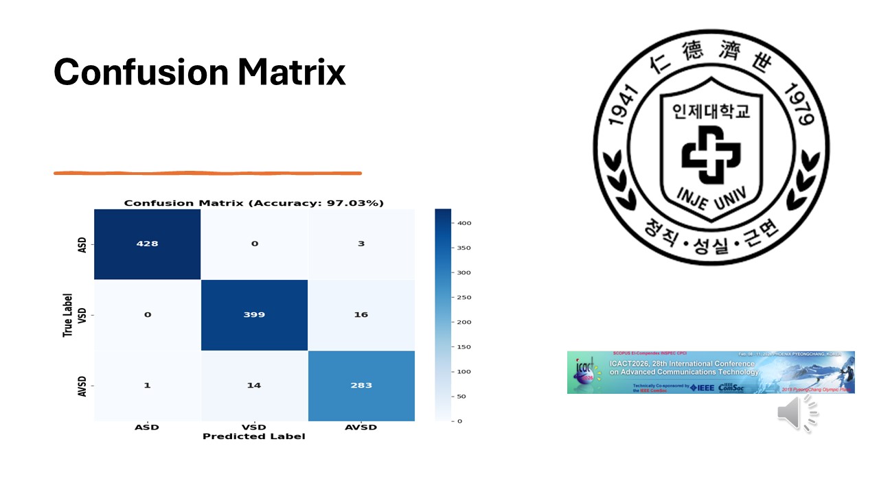 |
The confusion matrix shows the classification results across all three CHD classes.
The model demonstrates high accuracy with minimal misclassification between ASD, VSD, and AVSD categories. |
| ICACT20260266 Slide.17
[Big Slide]
|
Chrome Click!! |
 |
Grad-CAM++ visualization highlights regions that strongly influence model predictions. Warmer colors indicate areas of higher importance. These visualizations confirm that the model focuses on clinically relevant cardiac structures, improving trust and interpretability. |
| ICACT20260266 Slide.18
[Big Slide]
|
Chrome Click!! |
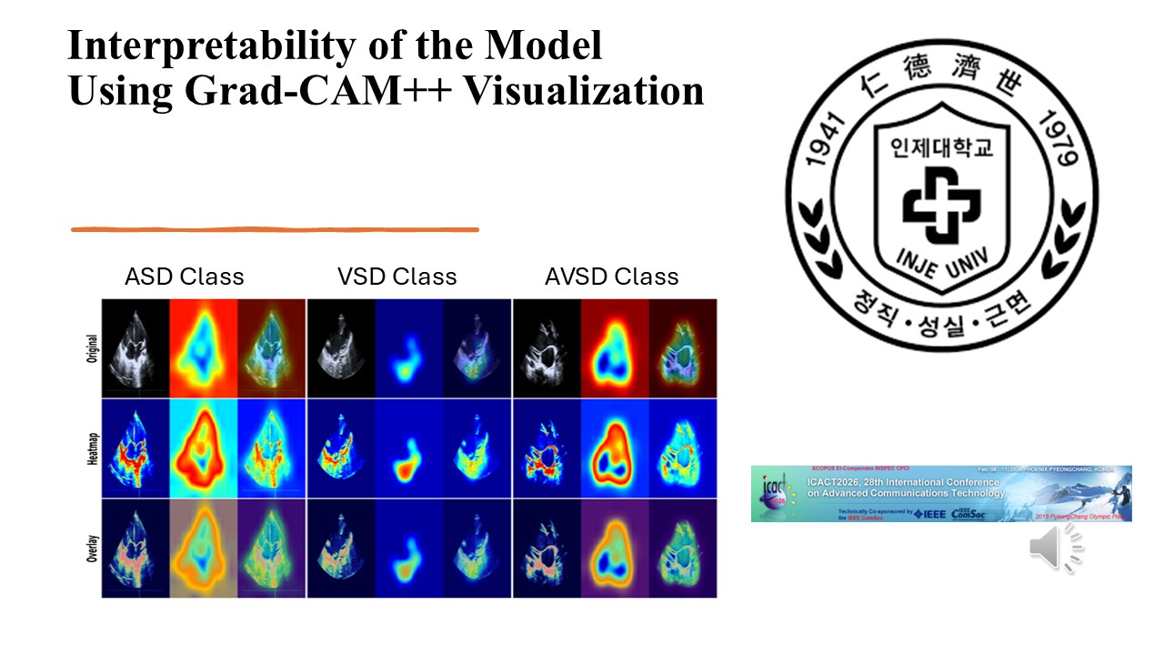 |
These examples show Grad-CAM++ heatmaps for ASD, VSD, and AVSD cases. The highlighted regions correspond to actual defect locations, demonstrating the clinical reliability of the proposed system. |
| ICACT20260266 Slide.19
[Big Slide]
|
Chrome Click!! |
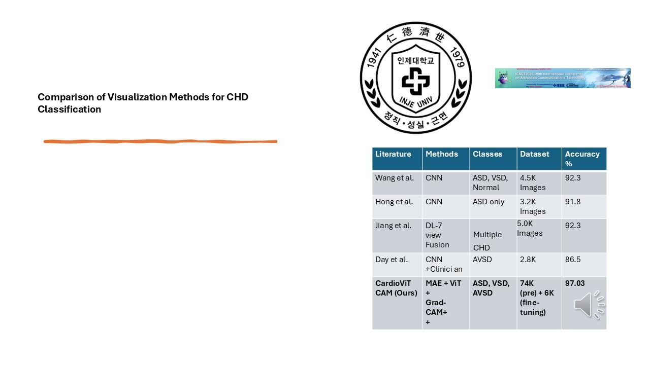 |
The comparative analysis of CHD classification models. In contrast to previous studies that focus on a single class or a limited number of defect types, the proposed MAE + ViT + Grad-CAM++ framework achieves superior accuracy across multiple CHD categories (i.e., ASD, VSD, and AVSD). Leveraging large-scale pretraining and domain-specific fine-tuning enhances feature representation and diagnostic precision. The accuracy achieved of 97.03% marks a significant improvement over existing CNN-based methodology. |
| ICACT20260266 Slide.20
[Big Slide]
|
Chrome Click!! |
 |
In conclusion, we proposed CardioViT-CAM, an interpretable deep learning framework for automated CHD classification.
The model integrates:
MAE self-supervised pretraining
Vision Transformer fine-tuning
Grad-CAM++ explainability
The system achieved 97.03% classification accuracy and shows strong potential for clinical decision support. |
| ICACT20260266 Slide.21
[Big Slide]
|
Chrome Click!! |
 |
Thank you. |